
Sat, Oct 3, 2026
[Archive]
Volume 34, Issue 3 (May & June 2026)
J Adv Med Biomed Res 2026, 34(3): 299-304 |
Back to browse issues page



Download citation:
BibTeX | RIS | EndNote | Medlars | ProCite | Reference Manager | RefWorks
Send citation to:


BibTeX | RIS | EndNote | Medlars | ProCite | Reference Manager | RefWorks
Send citation to:
Haji Zeinali A M, Bekravi M. Hypereosinophilic Syndrome Presented with Myocardial Infarction with Non-Obstructive Coronary Arteries: A case Report. J Adv Med Biomed Res 2026; 34 (3) :299-304
URL: http://journal.zums.ac.ir/article-1-7756-en.html
URL: http://journal.zums.ac.ir/article-1-7756-en.html


1- Department of interventional cardiology, Tehran Heart Center, Tehran University of Medical Sciences, Tehran, Iran
2- Department of interventional cardiology, Tehran Heart Center, Tehran University of Medical Sciences ,M_bekravi@yahoo.com
2- Department of interventional cardiology, Tehran Heart Center, Tehran University of Medical Sciences ,
Keywords: Myocardial Infarction with Non-Obstructive Coronary Arteries, Eosinophil, Hypereosinophilic syndrome.
Full-Text [PDF 448 kb]
(69 Downloads)
| Abstract (HTML) (522 Views)
Table 1. Timeline: Strategies and Findings.
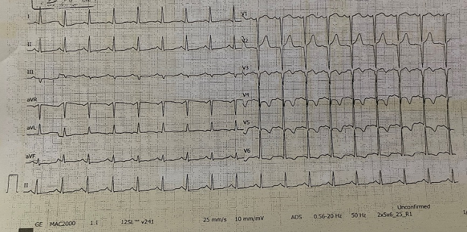
Figure 1. Electrocardiogram of the patient. (Prepared by Authors, 2026).
Note: Inverted T-wave in pericardial leads (V4-V6) is seen.
*Sinus tachycardia with a rate of 100-110, narrow QRS, biphasic T-wave in V3 and inverted T-wave in V4-V6. *
.jpg)
Figure 2. Tissue Doppler Imaging in Echocardiography. (Prepared by Authors, 2026).
Note: Blue arrow: Left ventricular (LV) clot , Red arrow: Pericardial effusion.
*The heart chambers are indicated within the figure. Moderate left ventricular hypertrophy (LVH) and a large sessile semi‑mobile mass in the left ventricular apex measuring 15 × 21 mm, with echogenicity similar to the adjacent myocardium. Mild pleural effusion was also noted.*
.jpg)
Figure 3. Cardiac magnetic resonance (CMR) imaging report. (Prepared by Authors, 2026).
Note: LV: Left ventrricular, RV: Right ventricular, LGE: late gadolinium enhancement.
Yellow arrow: LV clot, Blue arrow: Diffuse LGE enhancement.
*Diffuse late gadolinium enhancement (LGE) in the left ventricle, along with a well‑defined lobulated intracavitary mass at the LV apex (31 × 14 mm). The mass demonstrated iso‑ to high‑signal intensity on STIR, iso‑signal on BTFE, lack of enhancement on first‑pass perfusion and LGE sequences, and low‑signal intensity on T1 and T2 mapping, consistent with thrombus.
Full-Text: (39 Views)
Hypereosinophilic syndrome (HES) is characterized by persistent eosinophilia without any discernible underlying cause. The patient may exhibit no symptoms or may present with a disorder affecting multiple systems. Electrocardiography, echocardiography, and cardiac magnetic resonance imaging are commonly used methods to assess cardiac involvement in HES. Recognizing HES early can be difficult since the initial stage typically lacks noticeable clinical symptoms. Here, we report a 45-year-old man with HES that mimics the signs and symptoms of acute coronary syndrome. After ruling out other possible diagnoses, Electrocardiography revealed inverted T-waves in pericardial leads (V4–V6). Coronary angiography demonstrated non-obstructive coronary artery disease. Echocardiography showed moderate left ventricular (LV) hypertrophy, a lobulated apical LV mass, trivial tricuspid regurgitation, and mild pleural effusion. Cardiac MRI identified an apical LV mass with iso/high signal intensity on STIR sequences and no enhancement on late gadolinium enhancement (LGE), consistent with LV thrombus in the setting of HES. On admission, troponin level was 869 ng/ml. HES was confirmed, and the patient was treated with prednisolone (5 mg/kg daily) and apixaban (10 mg BID for 7 days, then 5 mg BID). Follow-up demonstrated a decrease in troponin to 351 ng/ml and complete resolution of the LV thrombus on echocardiography.
Keywords: Myocardial Infarction with Non-Obstructive Coronary Arteries, Eosinophil, Hypereosinophilic syndrome
1. Introduction
Hypereosinophilic syndrome is a rare hematopoietic disorder (1) characterized by persistent eosinophilia in the blood lasting more than 6 months, without any discernible underlying cause (2). It also includes an elevated number of eosinophils in the peripheral blood (blood eosinophils exceeding 1.5 × 10^9/l), along with organ dysfunction or damage attributed to eosinophil activity, after ruling out all other potential causes (3). HES can also lead to gastrointestinal, cardiovascular, neurological, and hematological complications. Among these, cardiac symptoms have the most critical impact on morbidity and mortality (4). Atherosclerotic coronary artery disease is the most common type of AMI; however, they also occur in people with non-obstructive coronary artery disease (MINOCA) (5). In the absence of angiography, visible pathology and lumen irregularities, including symptoms of atherosclerosis, thrombosis, or spasm, are called normal coronary angiograms (6). MINOCA comprises at least 1% - 12% of total myocardial infarction, and interest and awareness of MINOCA due to repeated use of coronary angiography has recently increased (7). Recognizing HES early can be difficult since the initial stage typically lacks noticeable clinical symptoms (8). Here, we report a case of HES that mimics ACS signs and symptoms.
2. Case presentation
A 45 –year- old non-smoker and non-alcoholic man who complained of retrosternal chest pain (CP) for 1 month arrived at Tehran Heart Center, Tehran, Iran. The characteristics of his CP were positional, exertional NYHA functional class (FC) III and aggravated during the last week, without similar past episodes and, he reported dyspnea on exertion (FC II). Also, he was suspected of having transient ischemic attack due to delirium and dizziness from 1 month ago, for which further investigations were normal. He had a history of intermittent fever, occasional chills, and asthma, for which he did not take any medication and exhibited no signs or symptoms indicative of a viral infection.
During the physical examination, the patient was conscious, and the initial vital signs were as follows: blood pressure, 110/70 mmHg; pulse rate, 110/min; respiratory rate, 17/min; and temperature, 37.9 °C. S1 and S2 were heard in cardiac auscultation and no murmur was found. During lung auscultation, decreased sounds in the bases of the lungs were heard. No chest deformity and no tenderness of chest and abdomen; were observed. The results of blood test at the arrival time was as below; white blood cell (WBC): 17600 µl, hemoglobin: 15.7 mg/dl, platelet; 25000 / µl, neutrophil: 27 %, eosinophil: 58.7 %, lymphocyte: 10 %, monocyte: 3 %, erythrocyte sedimentation rate: 8 mm/hr, C-reactive protein: 5 mg/L (normal range < 0.5) and quantitative troponin level was 869 ng/ml and negative Covid-19 PCR test. In further investigations, the WBC changed to 21000 and then 10000 /µl. Also, eosinophil count changed to 30 % and then 41 %. No rash, nodules, papules, Osler nodes, or Janeway lesions were seen on skin examination. The electrocardiogram (ECG) revealed sinus tachycardia with a rate of 100-110, normal axis, normal PR interval, narrow QRS and normal QRS progression, no ST segment deviation, biphasic T-wave in V3 and inverted T-wave in V4-V6, and normal QT (QTc= 408 ms). Because of high troponin level and abnormal ECG findings and his CP, coronary angiography (CAG) was performed, and the result was normal epicardial coronary artery (NECA). Moreover, a chest computed tomography (CT) scan indicated minimal bilateral pleural effusion and mild pericardial effusion. The echocardiography reported a left ventricular ejection fraction (LVEF) equal to 55-60 %, no regional wall motion abnormalities (RWMA), moderate left ventricular hypertrophy (LVH), a large sessile semi-mobile mass with echo density similar to adjacent myocardium in the left ventricular apex 15mm×21mm, normal right ventricular function, trivial tricuspid regurgitation (TR), and mild pleural effusion. Pulmonary artery pressure was 20 mmhg, reduced vertical separation minimum (RVSM) was 11 (Figure 1 and Figure 2).
Diffuse distribution of late gadolinium enhancement (LGE) in LV, well defined mass lobulated in some part of LV apex with measurement of 31×14mm iso-signal in balanced turbo field echo (BTFE), iso to high signal in short time inversion recovery (STIR), non-enhanced at first pass and LGE, low-signal in T1 and T2 mapping together with tissue characterization of mass and LV clot was observed in cardiac magnetic resonance (CMR) imaging report (Figure 3). Based on these findings, HES with an accompanyngLV clot was our first diagnosis. After these examinations,the patient was a candidate for cardiac biopsy and was evaluated for eosinophilic myocarditis, whichhe didn’t permit, so the procedure wasn’t done. To rull out other diagnoses fasciola antibody (Ab) (IgG), toxocara Ab and strongyloides Ab were checked and all were negative. There was no mutation in hotspot regions of platelet- derived growth factor alpha and beta receptor genes (PDGFRα and PDGFRβ), and all rheumatology tests, such as ANA, ANTI, DS, DNA, etc. were normal.
Finally, the diagnosis of HES was confirmed, and we prescribed Prednisolone 5 mg/kg daily and Apixaban 10 mg twice a day for 7 days, then 5 mg twice a day for LV clot. After 1month use of Apixaban the LV clot resolved as echocardiography demonstrated. The patient was discharged from the hospital after 5 days of hospitalization, and his troponin level decreased from 869 ng/ml in the initial evaluation to 351 ng/ml at the time of discharge (Table 1).
2. Case presentation
A 45 –year- old non-smoker and non-alcoholic man who complained of retrosternal chest pain (CP) for 1 month arrived at Tehran Heart Center, Tehran, Iran. The characteristics of his CP were positional, exertional NYHA functional class (FC) III and aggravated during the last week, without similar past episodes and, he reported dyspnea on exertion (FC II). Also, he was suspected of having transient ischemic attack due to delirium and dizziness from 1 month ago, for which further investigations were normal. He had a history of intermittent fever, occasional chills, and asthma, for which he did not take any medication and exhibited no signs or symptoms indicative of a viral infection.
During the physical examination, the patient was conscious, and the initial vital signs were as follows: blood pressure, 110/70 mmHg; pulse rate, 110/min; respiratory rate, 17/min; and temperature, 37.9 °C. S1 and S2 were heard in cardiac auscultation and no murmur was found. During lung auscultation, decreased sounds in the bases of the lungs were heard. No chest deformity and no tenderness of chest and abdomen; were observed. The results of blood test at the arrival time was as below; white blood cell (WBC): 17600 µl, hemoglobin: 15.7 mg/dl, platelet; 25000 / µl, neutrophil: 27 %, eosinophil: 58.7 %, lymphocyte: 10 %, monocyte: 3 %, erythrocyte sedimentation rate: 8 mm/hr, C-reactive protein: 5 mg/L (normal range < 0.5) and quantitative troponin level was 869 ng/ml and negative Covid-19 PCR test. In further investigations, the WBC changed to 21000 and then 10000 /µl. Also, eosinophil count changed to 30 % and then 41 %. No rash, nodules, papules, Osler nodes, or Janeway lesions were seen on skin examination. The electrocardiogram (ECG) revealed sinus tachycardia with a rate of 100-110, normal axis, normal PR interval, narrow QRS and normal QRS progression, no ST segment deviation, biphasic T-wave in V3 and inverted T-wave in V4-V6, and normal QT (QTc= 408 ms). Because of high troponin level and abnormal ECG findings and his CP, coronary angiography (CAG) was performed, and the result was normal epicardial coronary artery (NECA). Moreover, a chest computed tomography (CT) scan indicated minimal bilateral pleural effusion and mild pericardial effusion. The echocardiography reported a left ventricular ejection fraction (LVEF) equal to 55-60 %, no regional wall motion abnormalities (RWMA), moderate left ventricular hypertrophy (LVH), a large sessile semi-mobile mass with echo density similar to adjacent myocardium in the left ventricular apex 15mm×21mm, normal right ventricular function, trivial tricuspid regurgitation (TR), and mild pleural effusion. Pulmonary artery pressure was 20 mmhg, reduced vertical separation minimum (RVSM) was 11 (Figure 1 and Figure 2).
Diffuse distribution of late gadolinium enhancement (LGE) in LV, well defined mass lobulated in some part of LV apex with measurement of 31×14mm iso-signal in balanced turbo field echo (BTFE), iso to high signal in short time inversion recovery (STIR), non-enhanced at first pass and LGE, low-signal in T1 and T2 mapping together with tissue characterization of mass and LV clot was observed in cardiac magnetic resonance (CMR) imaging report (Figure 3). Based on these findings, HES with an accompanyngLV clot was our first diagnosis. After these examinations,the patient was a candidate for cardiac biopsy and was evaluated for eosinophilic myocarditis, whichhe didn’t permit, so the procedure wasn’t done. To rull out other diagnoses fasciola antibody (Ab) (IgG), toxocara Ab and strongyloides Ab were checked and all were negative. There was no mutation in hotspot regions of platelet- derived growth factor alpha and beta receptor genes (PDGFRα and PDGFRβ), and all rheumatology tests, such as ANA, ANTI, DS, DNA, etc. were normal.
Finally, the diagnosis of HES was confirmed, and we prescribed Prednisolone 5 mg/kg daily and Apixaban 10 mg twice a day for 7 days, then 5 mg twice a day for LV clot. After 1month use of Apixaban the LV clot resolved as echocardiography demonstrated. The patient was discharged from the hospital after 5 days of hospitalization, and his troponin level decreased from 869 ng/ml in the initial evaluation to 351 ng/ml at the time of discharge (Table 1).
Table 1. Timeline: Strategies and Findings.
| Strategies (Clinical Actions & Diagnostics) | Trends and Findings (Clinical Changes & Results) |
| Patient presented with chest pain and initial assessment | Retrosternal chest pain (NYHA class III), exertional dyspnea (class II), no prior similar episodes |
| Physical examination and vital signs | BP: 110/70 mmHg, HR: 110 bpm, Temp: 37.9°C, decreased breath sounds at lung bases |
| Initial blood tests | WBC: 17,600/µl, Platelet: 250,000/µl, Eosinophil: 58.7%, CRP: 5 mg/L, Troponin: 869 ng/ml, COVID-19 PCR negative |
| Follow-up blood tests | WBC rose to 21,000 then dropped to 10,000/µl; eosinophil levels changed to 30% then 41% |
| Electrocardiogram (ECG) | Inverted T-waves in pericardial leads (V4–V6) |
| Coronary angiography (CAG) | Non-obstructive coronary artery disease |
| Chest CT scan | Mild bilateral pleural effusion and pericardial effusion |
| Echocardiography | LVEF: 55–60%, moderate LV hypertrophy, apical LV mass (15×21 mm), trivial TR, mild pleural effusion |
| Cardiac MRI | Lobulated LV apical mass (31×14 mm), iso/high signal in STIR, no enhancement in LGE, consistent with LV clot and HES |
| Differential diagnosis workup | Negative for parasitic antibodies, normal rheumatologic tests, no PDGFRα/β mutations |
| Treatment initiated | Prednisolone 5 mg/kg daily, Apixaban 10 mg BID for 7 days than 5 mg BID |
| Follow-up and discharge | Troponin decreased to 351 ng/ml, LV clot resolved on echo, discharged after 5 days |
Figure 1. Electrocardiogram of the patient. (Prepared by Authors, 2026).
Note: Inverted T-wave in pericardial leads (V4-V6) is seen.
*Sinus tachycardia with a rate of 100-110, narrow QRS, biphasic T-wave in V3 and inverted T-wave in V4-V6. *
Figure 2. Tissue Doppler Imaging in Echocardiography. (Prepared by Authors, 2026).
Note: Blue arrow: Left ventricular (LV) clot , Red arrow: Pericardial effusion.
*The heart chambers are indicated within the figure. Moderate left ventricular hypertrophy (LVH) and a large sessile semi‑mobile mass in the left ventricular apex measuring 15 × 21 mm, with echogenicity similar to the adjacent myocardium. Mild pleural effusion was also noted.*
Figure 3. Cardiac magnetic resonance (CMR) imaging report. (Prepared by Authors, 2026).
Note: LV: Left ventrricular, RV: Right ventricular, LGE: late gadolinium enhancement.
Yellow arrow: LV clot, Blue arrow: Diffuse LGE enhancement.
*Diffuse late gadolinium enhancement (LGE) in the left ventricle, along with a well‑defined lobulated intracavitary mass at the LV apex (31 × 14 mm). The mass demonstrated iso‑ to high‑signal intensity on STIR, iso‑signal on BTFE, lack of enhancement on first‑pass perfusion and LGE sequences, and low‑signal intensity on T1 and T2 mapping, consistent with thrombus.
3. Discussion
Cardiac presentation of HES includes asymptomatic myocardial involvement, ischemic events, stroke, heart failure, fatal arrhythmias, and sudden cardiac death. In our case, the patient presented with cardiac symptoms; after cardiac evaluation ACS was ruled out and the cardiac manifestation represents myocarditis. Therefore, other investigations were also carried out, and finally, an HES diagnosis was confirmed. As reported previously, HES can lead to cardiovascular complications, and cardiac symptoms have the most critical impact on morbidity and mortality of patients (4). HES must be distinguished from other reasons for hypereosinophilia, including parasitic infections or malignancies (9). HES most commonly affect middle-aged males. In a 2024 case series from the Cleveland Clinic, the majority of patients with eosinophilic myocarditis were male, with a median age of 43 years (10). Our patient, a 45-year-old male, aligns with these demographic patterns. Cardiac involvement in HES often presents with chest pain, dyspnea, and fatigue. In the 2023 EHJ Case Report, the patient experienced progressive dyspnea and weight loss due to cardiac cachexia (11). Another 2024 report in Frontiers in Immunology described eosinophilic myocarditis presenting with chest pain, dizziness, and signs of heart failure (12). Our patient’s exertional chest pain (NYHA III), dyspnea (NYHA II), and transient neurological symptoms are consistent with these findings. Electrocardiographic abnormalities in HES are typically non-specific. A 2024 review in ABC Cardiology noted that T-wave inversions and ST-segment changes may occur due to eosinophilic infiltration of the myocardium (13). Our patient’s T-wave inversion in leads V4–V6 supports myocardial involvement and is consistent with prior reports. Cardiac MRI is critical for evaluating eosinophilic myocarditis. A study identified diffuse late gadolinium enhancement (LGE), iso- to high-signal intensity on STIR, and low signal on T1/T2 mapping as typical features of HES-related myocardial damage (14). Our patient’s MRI showed all these features, including a lobulated LV apical mass consistent with thrombus, confirming the diagnosis. ECG and echocardiography are commonly used methods to assess cardiac involvement in HES. Non-specific ECG changes, such as ST- segment and T-wave changes or LVH, may be observed. The most significant echocardiographic findings in HES include thrombosis in the left or right ventricle and endomyocardial thickening (15). We found T-inversion in the ECG and LVH in echocardiography and a high troponin level during assessment of our case, which made us close to diagnosing HES. Increasingly, we found trivial TR in patient's echocardiography. Treatment strategies for HES vary based on its subtype and severity (including cardiac, central nervous system, or thrombotic involvement), clinical progression (continuous or relapsing/remitting), and patient's characteristics (such as age and potential comorbidities). Eosinophils are activated by cytokines such as IL-5, IL-3, and GM-CSF, which promote their survival and recruitment to tissues. Once activated, eosinophils migrate into the myocardium, where they release cytotoxic granules. These granules cause direct myocardial damage, endothelial injury, and promote thrombogenesis (16).
Corticosteroid therapy has been suggested for the treatment of HES. Therefore, we prescribed prednisolone to our case, which seemed to be effective. According to Klion et al., systemic corticosteroids like prednisolone are effective in reducing eosinophilia and preventing organ damage in HES (17). In a 2023 Case Report, a patient with HES and cardiac cachexia was treated with corticosteroids and anticoagulation (initial LMWH followed by warfarin) (11). Our case is notable for using apixaban instead of warfarin, reflecting a modern approach supported by emerging evidence. One study used cytotoxic Agents like hydroxyurea in steroid-refractory cases or when rapid eosinophil reduction is needed. It inhibits DNA synthesis and reduces eosinophil proliferation (11).
4. Conclusion
HES is an uncommon condition, and limited robust epidemiological data are available. In this case, we concluded that HES could be considered as a differential diagnosis of ACS and myocarditis if a patient presents with CP and a high troponin level after ruling out ACS.
5. Declarations
5.1 Acknowledgments
We would like to thank the patient for participating in this case report.
5.2 Ethical Considerations
The patient provided informed consent for inclusion in this case report.
5.3 Authors' Contributions
Ali Mohammad Haji Zeinali: Contributed to the study design, data collection, analysis and writing of the manuscript. Mahzad Bekravi: Manuscript drafting. The final manuscript was read and approved by all of the authors.
5.4 Conflict of Interest
The authors declared no conflicts of interest.
5.5 Fund or Financial Support
This research received no specific grant from any funding agency in the public, commercial, or not- for- profit sector.
5.6 Using Artificial Intelligence Tools (AI Tools)
The authors were not utilized AI Tools.
Cardiac presentation of HES includes asymptomatic myocardial involvement, ischemic events, stroke, heart failure, fatal arrhythmias, and sudden cardiac death. In our case, the patient presented with cardiac symptoms; after cardiac evaluation ACS was ruled out and the cardiac manifestation represents myocarditis. Therefore, other investigations were also carried out, and finally, an HES diagnosis was confirmed. As reported previously, HES can lead to cardiovascular complications, and cardiac symptoms have the most critical impact on morbidity and mortality of patients (4). HES must be distinguished from other reasons for hypereosinophilia, including parasitic infections or malignancies (9). HES most commonly affect middle-aged males. In a 2024 case series from the Cleveland Clinic, the majority of patients with eosinophilic myocarditis were male, with a median age of 43 years (10). Our patient, a 45-year-old male, aligns with these demographic patterns. Cardiac involvement in HES often presents with chest pain, dyspnea, and fatigue. In the 2023 EHJ Case Report, the patient experienced progressive dyspnea and weight loss due to cardiac cachexia (11). Another 2024 report in Frontiers in Immunology described eosinophilic myocarditis presenting with chest pain, dizziness, and signs of heart failure (12). Our patient’s exertional chest pain (NYHA III), dyspnea (NYHA II), and transient neurological symptoms are consistent with these findings. Electrocardiographic abnormalities in HES are typically non-specific. A 2024 review in ABC Cardiology noted that T-wave inversions and ST-segment changes may occur due to eosinophilic infiltration of the myocardium (13). Our patient’s T-wave inversion in leads V4–V6 supports myocardial involvement and is consistent with prior reports. Cardiac MRI is critical for evaluating eosinophilic myocarditis. A study identified diffuse late gadolinium enhancement (LGE), iso- to high-signal intensity on STIR, and low signal on T1/T2 mapping as typical features of HES-related myocardial damage (14). Our patient’s MRI showed all these features, including a lobulated LV apical mass consistent with thrombus, confirming the diagnosis. ECG and echocardiography are commonly used methods to assess cardiac involvement in HES. Non-specific ECG changes, such as ST- segment and T-wave changes or LVH, may be observed. The most significant echocardiographic findings in HES include thrombosis in the left or right ventricle and endomyocardial thickening (15). We found T-inversion in the ECG and LVH in echocardiography and a high troponin level during assessment of our case, which made us close to diagnosing HES. Increasingly, we found trivial TR in patient's echocardiography. Treatment strategies for HES vary based on its subtype and severity (including cardiac, central nervous system, or thrombotic involvement), clinical progression (continuous or relapsing/remitting), and patient's characteristics (such as age and potential comorbidities). Eosinophils are activated by cytokines such as IL-5, IL-3, and GM-CSF, which promote their survival and recruitment to tissues. Once activated, eosinophils migrate into the myocardium, where they release cytotoxic granules. These granules cause direct myocardial damage, endothelial injury, and promote thrombogenesis (16).
Corticosteroid therapy has been suggested for the treatment of HES. Therefore, we prescribed prednisolone to our case, which seemed to be effective. According to Klion et al., systemic corticosteroids like prednisolone are effective in reducing eosinophilia and preventing organ damage in HES (17). In a 2023 Case Report, a patient with HES and cardiac cachexia was treated with corticosteroids and anticoagulation (initial LMWH followed by warfarin) (11). Our case is notable for using apixaban instead of warfarin, reflecting a modern approach supported by emerging evidence. One study used cytotoxic Agents like hydroxyurea in steroid-refractory cases or when rapid eosinophil reduction is needed. It inhibits DNA synthesis and reduces eosinophil proliferation (11).
4. Conclusion
HES is an uncommon condition, and limited robust epidemiological data are available. In this case, we concluded that HES could be considered as a differential diagnosis of ACS and myocarditis if a patient presents with CP and a high troponin level after ruling out ACS.
5. Declarations
5.1 Acknowledgments
We would like to thank the patient for participating in this case report.
5.2 Ethical Considerations
The patient provided informed consent for inclusion in this case report.
5.3 Authors' Contributions
Ali Mohammad Haji Zeinali: Contributed to the study design, data collection, analysis and writing of the manuscript. Mahzad Bekravi: Manuscript drafting. The final manuscript was read and approved by all of the authors.
5.4 Conflict of Interest
The authors declared no conflicts of interest.
5.5 Fund or Financial Support
This research received no specific grant from any funding agency in the public, commercial, or not- for- profit sector.
5.6 Using Artificial Intelligence Tools (AI Tools)
The authors were not utilized AI Tools.
Type of Study: Case Report Article |
Subject:
Clinical Medicine
Received: 2026/02/10 | Accepted: 2026/06/22 | Published: 2026/06/29
Received: 2026/02/10 | Accepted: 2026/06/22 | Published: 2026/06/29
References
1. Ryan D, Inamullah O, El Husseini N, Wang E, Selim MA, Feng W. The hypereosinophilic syndrome-an unusual cause of myocarditis and cardioembolic strokes. Am J Med Sci. 2022; 364(5): 661-8. [DOI:10.1016/j.amjms.2022.05.019] [PMID]
2. Tahouri T, Mahdavi M, Rezaei-Kalantari K, Shahzadi H. Irreversible coronary aneurysm presenting as acute coronary syndrome in a child with hypereosinophilic syndrome: a case report. Cardiol Young. 2021; 31(5): 845-7. [DOI:10.1017/S1047951120004862] [PMID]
3. Kleinfeldt T, Nienaber C, Kische S, Akin I, Turan R, Körber T, et al. Cardiac manifestation of the hypereosinophilic syndrome: new insights. Clin Res Cardiol. 2010; 99: 419-27. [DOI:10.1007/s00392-010-0144-8] [PMID]
4. Bondue A, Carpentier C, Roufosse F. Hypereosinophilic syndrome: considerations for the cardiologist. Heart. 2022; 108(3): 164-71. [DOI:10.1136/heartjnl-2020-317202] [PMID]
5. Helbig G, Kyrcz‐Krzemień S. Cessation of imatinib mesylate may lead to sustained hematologic and molecular remission in FIP1L1‐PDGFRA‐mutated hypereosinophilic syndrome. Am J Hematol. 2014; 89(1): 115-. [DOI:10.1002/ajh.23588] [PMID]
6. Menzella F, Lusuardi M, Galeone C, Taddei S, Zucchi L. Profile of anti-IL-5 mAb mepolizumab in the treatment of severe refractory asthma and hypereosinophilic diseases. J Asthma Allergy. 2015: 105-14. [DOI:10.2147/JAA.S40244] [PMID] [PMCID]
7. Kolbeck R, Kozhich A, Koike M, Peng L, Andersson CK, Damschroder MM, et al. MEDI-563, a humanized anti-IL-5 receptor α mAb with enhanced antibody-dependent cell-mediated cytotoxicity function. J Allergy Clin Immunol. 2010; 125(6): 1344-53. e2. [DOI:10.1016/j.jaci.2010.04.004] [PMID]
8. Dzelebdzic S, Sasaki N, Welch E, Muniz JC. Hypereosinophilic syndrome with advanced-stage loeffler endocarditis. CASE: Cardiovasc Imag Case Report. 2022; 6(4): 191. [DOI:10.1016/j.case.2022.02.004] [PMID] [PMCID]
9. Wang SA. The diagnostic work-up of hypereosinophilia. Pathobiol. 2019; 86(1): 39-52. [DOI:10.1159/000489341] [PMID]
10. Reeder M, Okushi Y, Vega SLP, Prasad R, Grimm RA, Griffin BP, et al. The Cleveland Clinic experience of eosinophilic myocarditis in the setting of hypereosinophilic syndrome: demographics, cardiac imaging, and outcomes. Cardiovasc Diag Ther. 2024; 14(6): 1122. [DOI:10.21037/cdt-24-347] [PMID] [PMCID]
11. Teringová E, Penz P, Mištinová JP, Orban M. Hypereosinophilic syndrome presenting with progressive cardiac cachexia: a case report. Europ Heart J Case Report. 2023; 7(7): ytad280. [DOI:10.1093/ehjcr/ytad280] [PMID] [PMCID]
12. Sharma S, Desai S, Leoni J, Paghdar S, Ruiz J, Goswami R. Case report: eosinophilic myocarditis in hypereosinophilic syndrome: a journey to heart transplantation. Front Immunol. 2024; 15: 1418665. [DOI:10.3389/fimmu.2024.1418665] [PMID] [PMCID]
13. Hotta VT, Nastari RR, Oishi GdSL, Kayano AE, Oliveira JAG, Rocha RG, et al. Cardiac abnormalities in hypereosinophilic syndromes. Arquiv Brasil Cardiol. 2024; 121: e20240190. [DOI:10.36660/abc.20240190i]
14. Merten C, Beurich H, Zachow D, Arndt F, Moosig F, Richardt G. Cardiac involvement in hypereosinophilic syndromes detected by cardiac magnetic resonance imaging. J Cardiovasc Magnet Reson. 2015; 17. [DOI:10.1186/1532-429X-17-S1-Q75] [PMCID]
15. Ogbogu PU, Rosing DR, Horne III MK. Cardiovascular manifestations of hypereosinophilic syndromes. Immunol Allergy Clin North Am. 2007; 27(3): 457-75. [DOI:10.1016/j.iac.2007.07.001] [PMID] [PMCID]
16. Piccirillo F, Mastroberardino S, Nafisio V, Fiorentino M, Segreti A, Nusca A, et al. Eosinophilic myocarditis: from bench to bedside. Biomed. 2024; 12(3): 656. [DOI:10.3390/biomedicines12030656] [PMID] [PMCID]
17. Klion A. Hypereosinophilic syndrome: approach to treatment in the era of precision medicine. Hematology 2014, the American Society of Hematology Education Program Book. 2018; 2018(1): 326-31. [DOI:10.1182/asheducation-2018.1.326] [PMID] [PMCID]
Send email to the article author
| Rights and permissions | |
 |
This work is licensed under a Creative Commons Attribution-NonCommercial 4.0 International License. |
Copyright Policy
Journal of Advances in Medical and Biomedical Research by Zanjan University of Medical Sciences is licensed under CC BY-NC 4.0