
Thu, Sep 10, 2026
[Archive]
Volume 34, Issue 2 (March & April 2026)
J Adv Med Biomed Res 2026, 34(2): 120-133 |
Back to browse issues page


Download citation:
BibTeX | RIS | EndNote | Medlars | ProCite | Reference Manager | RefWorks
Send citation to:


BibTeX | RIS | EndNote | Medlars | ProCite | Reference Manager | RefWorks
Send citation to:
Azizi Jalilian F, Aghaei S, Heidarzadi K, Shokri S, Amini R, Pakzad I, et al . Natural Antimicrobial and Wound-Healing Properties of Grape and Raisin Extracts: Insights from In Vitro Assays. J Adv Med Biomed Res 2026; 34 (2) :120-133
URL: http://journal.zums.ac.ir/article-1-7809-en.html
URL: http://journal.zums.ac.ir/article-1-7809-en.html
Farid Azizi Jalilian1 

 , Safoora Aghaei2 , Kobra Heidarzadi3 , Somayeh Shokri4 , Razieh Amini5 , Iraj Pakzad6 , Morovat Taherikalani7 , Shahab Mahmoudvand8 , Nastaran Ansari4 , Zahra Ramezannia4 , Amir Keshavarzi9 , Gholamreza Kalvandi *10
, Safoora Aghaei2 , Kobra Heidarzadi3 , Somayeh Shokri4 , Razieh Amini5 , Iraj Pakzad6 , Morovat Taherikalani7 , Shahab Mahmoudvand8 , Nastaran Ansari4 , Zahra Ramezannia4 , Amir Keshavarzi9 , Gholamreza Kalvandi *10
1- Department of Medical Microbiology, Faculty of Medicine, Ilam University of Medical Sciences, Ilam, Iran. & Research Center for Molecular Medicine, Institute of Cancer, Hamadan University of Medical Sciences, Hamadan, Iran
2- Student Research Center, Ilam University of Medical Sciences, Ilam, Iran
3- Department of Medical Microbiology, Faculty of Medicine, Ilam University of Medical Sciences, Ilam, Iran.
4- Department of Virology, School of Medicine, Hamadan University of Medical Sciences, Hamadan, Iran
5- Research Center for Molecular Medicine, Institute of Cancer, Hamadan University of Medical Sciences, Hamadan, Iran
6- Department of Medical Microbiology, Faculty of Medicine, Ilam University of Medical Sciences, Ilam, Iran
7- Department of Microbiology, School of Medicine, Lorestan University of Medical Sciences, Khorramabad, Iran
8- Department of Virology, School of Medicine, Hamadan University of Medical Sciences, Hamadan, Iran & Research Center for Molecular Medicine, Institute of Cancer, Hamadan University of Medical Sciences, Hamadan, Iran
9- Behavioral Disorders and Substance Abuse Research Center, Hamadan University of Medical Sciences, Hamadan, Iran
10- Department of Pediatrics, School of Medicine, Hamadan University of Medical Sciences, Hamadan, Iran
2- Student Research Center, Ilam University of Medical Sciences, Ilam, Iran
3- Department of Medical Microbiology, Faculty of Medicine, Ilam University of Medical Sciences, Ilam, Iran.
4- Department of Virology, School of Medicine, Hamadan University of Medical Sciences, Hamadan, Iran
5- Research Center for Molecular Medicine, Institute of Cancer, Hamadan University of Medical Sciences, Hamadan, Iran
6- Department of Medical Microbiology, Faculty of Medicine, Ilam University of Medical Sciences, Ilam, Iran
7- Department of Microbiology, School of Medicine, Lorestan University of Medical Sciences, Khorramabad, Iran
8- Department of Virology, School of Medicine, Hamadan University of Medical Sciences, Hamadan, Iran & Research Center for Molecular Medicine, Institute of Cancer, Hamadan University of Medical Sciences, Hamadan, Iran
9- Behavioral Disorders and Substance Abuse Research Center, Hamadan University of Medical Sciences, Hamadan, Iran
10- Department of Pediatrics, School of Medicine, Hamadan University of Medical Sciences, Hamadan, Iran
Keywords: Antibacterial activity, Biofilm inhibition, Grape extracts, Natural compounds, Wound healing
Full-Text [PDF 752 kb]
(152 Downloads)
| Abstract (HTML) (754 Views)
Full-Text: (4 Views)
ABSTRACT
Background & Objective: This study examines the in vitro antimicrobial properties of black and yellow grape juice and raisin extracts against 12 Gram-positive and Gram-negative bacterial strains, as well as five Candida species.
Materials & Methods: Cell viability was assessed by MTT assay, antibacterial activity by disk diffusion, MIC and MBC tests, biofilm inhibition by a microtiter plate method, and wound healing potential by a scratch assay.
Results: MTT assay showed that black and yellow grape juices and yellow raisins were non-toxic up to 30 µg/mL, while black raisins were safe only up to 15 µg/mL. Disk diffusion demonstrated antibacterial activity of all extracts against most tested bacterial except A. baumannii and Methicillin-resistant S. aureus. The strongest inhibition zones were observed for P. aeruginosa, Enterococcus, S. epidermidis, L. monocytogenes, and S. maltophilia, varying by extract type, while E. coli and E. coli O157 showed the lowest sensitivity. None of the extracts showed antifungal activity against Candida species. The lowest MIC observed for the yellow and black raisin extracts was 15 µg/mL against Enterococcus and S. maltophilia. In the yellow and black grape juice, the lowest MIC against S. epidermidis, Enterococcus, and S. maltophilia was 30 µg/mL. Black grape juice showed the greatest inhibition of P. aeruginosa biofilm formation. Both yellow and black grape juices promoted wound healing.
Conclusion: Grape extracts may serve as promising natural sources for the development of novel antibacterial agents against the tested Gram-positive and Gram-negative bacteria.
Keywords: Antibacterial activity, Biofilm inhibition, Grape extracts, Natural compounds, Wound healing
To date, multiple antimicrobials have been developed, which are essential drugs used to treat mild to severe infections (1). While these antimicrobials have played a pivotal role in medical development, a growing and alarming concern in 21st-century medicine is the increasing resistance to antibiotics, which has emerged as one of the most significant problems in public health (2). This poses a threat to the community and hospitals in the treatment of common infections (3). Annual deaths worldwide due to antimicrobial resistance (AMR) reveal at least 1.27 million deaths per year and could rise even higher, to 10 million deaths a year by 2050 (4) . Additionally, multi-drug resistance (MDR) of pathogenic bacterial strains to modern antibiotics has developed over the past decades (5), accounting for hundreds of thousands of infections and significant mortality (6). Therefore, the search for alternative treatments, including herbal drugs, has become a focus of research (7).
Today, in many parts of the developing world, between 70% and 95% of people continue to rely on traditional medicine (TM), and many countries have integrated herbal medicine into their mainstream healthcare systems through regulations (8). Several studies have investigated the effectiveness of plant extracts and secondary metabolites that are endowed with antimicrobial properties, either alone or in combination with existing drugs (9-12). The low cost, biocompatibility, and effectiveness of herbal drugs make them an attractive source for the development of antimicrobial agents that enhance the biological activity of existing drugs (13, 14). Examples of medicinal plants with broad-spectrum antimicrobial activities include Allium sativum (garlic), Azadirachta indica (neem), Curcuma longa (15).
Grapes (Vitis vinifera L.) and their derivatives, including juice, pomace, and raisins, are rich sources of polyphenols such as proanthocyanidins, flavonoids, and resveratrol. These compounds exhibit antioxidant, anti-inflammatory, anticancer, antibacterial, and antifungal activities. Previous studies have shown that grape polyphenols can interact with bacterial cell walls, alter membrane permeability, inhibit microbial enzymes, and reduce biofilm formation (16). Therefore, evaluating the antimicrobial effects of grape and raisin extracts provides insights into their potential as natural alternatives or adjunctive therapies for treating infectious diseases. Furthermore, in addition to their antimicrobial activity, grape extracts have been reported to promote wound healing by enhancing fibroblast proliferation, stimulating angiogenesis, and modulating inflammatory responses. This dual functionality makes grape and raisin extracts promising candidates for applications in both infection control and tissue repair (17).
This study aimed to evaluate the effects of black and yellow grape juices, as well as raisin extracts, on wound healing and their antibacterial and antifungal activities. Additionally, the cytotoxicity of these extracts in fibroblast cells and their effects at safe concentrations on standard bacterial strains, clinical isolates, biofilm formation, and wound healing were also assessed.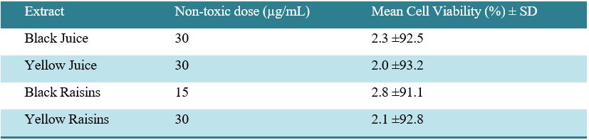
Disk diffusion tests were performed for all four types of extracts at concentrations of 7.5, 15, and 30 µg/mL. All bacteria, except A. baumannii and Methicillin-resistant S. aureus, showed zones of inhibition. The maximum diameter growth of inhibition zone in millimeters (mm) for black raisin extracts in the concentration of 30 µg/mL belonged to (P. aeruginosa~25 mm), (L. monocytogenes~24 mm), (Enterococcus~22 mm), (S. maltophilia~21 mm), (S. epidermidis~20 mm), for yellow raisin extracts maximum growth was observed in (Enterococcus~30mm), (P. aeruginosa~28 mm), (S. epidermidis~24 mm), (L. monocytogenes~23 mm), regarding the yellow juice the maximum rate was visible in (P. aeruginosa and Enterococcus~29 mm), (L. monocytogenes~25 mm), (S. maltophilia~23 mm), and finally for black grape extract the maximum rate was dominant in (P. aeruginosa~26 mm) and (Enterococcus and S. epidermidis~20 mm).
The minimum growth rate, as indicated by the inhibition zone diameter, was observed for all four extracts at different concentrations in E. coli and E. coli O157. At a concentration of 7.5 µg/mL, the three extracts (yellow raisin, black raisin, and yellow raisin juice) showed no inhibitory effect against E. coli O157. The highest average sensitivity among the bacterial strains was observed in Enterococcus toward the yellow raisin extracts, and the lowest sensitivity belonged to E. coli O157 in yellow grape juice (Figure 1). As shown in Figure 2, among the tested extracts, black raisin and yellow grape juice exhibited the highest overall inhibitory effects. Among the bacterial strains, P. aeruginosa and Enterococcus were the most susceptible to all extracts. The zone of inhibition produced by two bacterial strains, S. maltophilia, and E. coli, are shown in Figure 3.
The results of Figure 4 show the relationship between increased concentration and the growth inhibition zone.
The anti-fungal activity of the extracts against C.glabrata,
C.parapsilosis, C.albicans, C. tropicalis, and C.krusei was not detected; therefore, the tests were continued only with the sensitive bacterial strains.
3.3 MIC and MBC
The MIC values of all the plant extracts against the microorganisms are shown in Table 2. The concentrations of the plant extracts considered for the MIC assay were 7.5, 15, 30, 60, 120, and 240 µg/mL. These extracts exhibited antibacterial activity against the tested microorganisms, although the degree of activity varied among the bacterial species. The lowest MIC (highest antibacterial activity) for yellow and black raisin extracts was 15 µg/mL against Enterococcus, and S. maltophilia. In the yellow and black grape juice, the lowest MIC for S. epidermidis, S. maltophilia, and Enterococcus was observed at a concentration of 30 µg/mL. The highest MIC value (240 µg/mL), indicating the lowest antibacterial activity, was observed for both yellow and black raisin extracts against E. coli O157.
Results from the MBC assay supported the data obtained from the disk diffusion assay and MIC assay (Table 3).
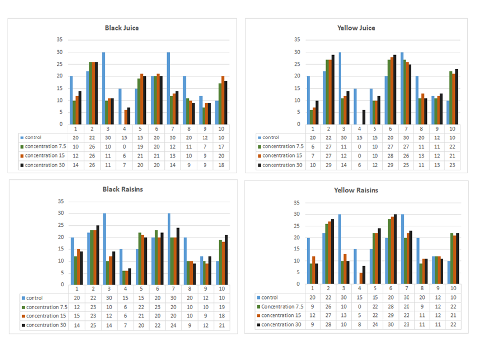
Figure 1. Growth rate of inhibition zone diameter. Bacteria name: 1. Helicobacter pylori, 2. Pseudomonas aeruginosa, 3. Escherichia coli, 4. Escherichia coli O157, 5. Staph epidermidis, 6. Enterococcus, 7. Listeria monocytogenes, 8. Methicillin sensitive staph aureus, 9. Salmonella typhimurium, 10. Stenotrophomonas maltophilia. (Prepared by Authors, 2026).
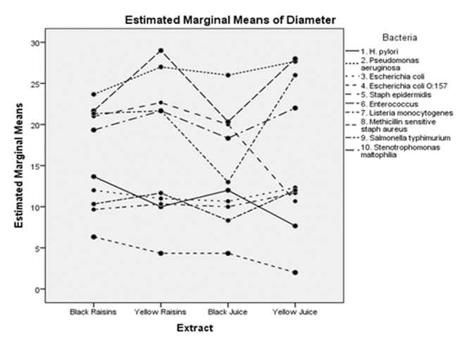
Figure 2. Estimated marginal means of inhibition zone diameters for 10 bacterial strains exposed to four grape-derived extracts (Black Raisins, Yellow Raisins, Black Juice, and Yellow Juice. (Prepared by Authors, 2026).
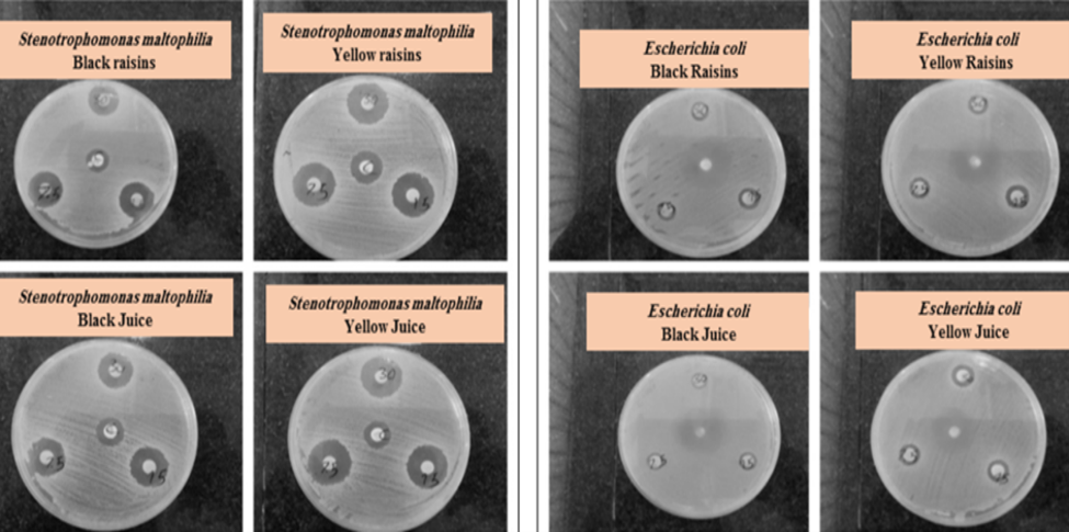
Figure 3. The diameter of inhibition zone in Stenotrophomonas maltophilia and E.coli. Left box bacteria: Stenotrophomonas maltophilia, Right box bacteria: Escherichia coli. First row in each box from left to right: Black raisins, raisins; Second row in each box from left to right: Black juice, yellow juice. (Prepared by Authors, 2026).
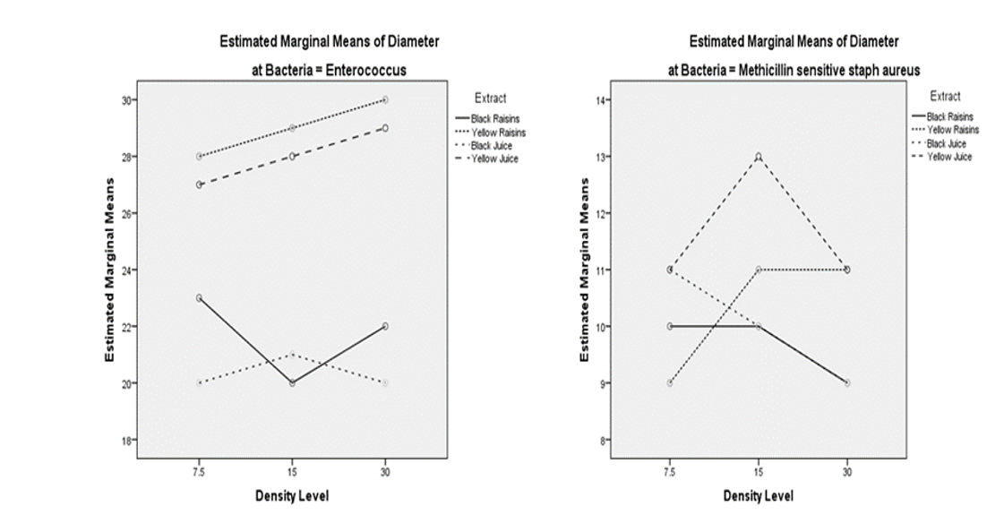
Figure 4. Relationship between increased concentration and growth inhibition zone. (Prepared by Authors, 2026).
Table 2. Minimum inhibitory concentrations of the plant extracts against the pathogenic organisms
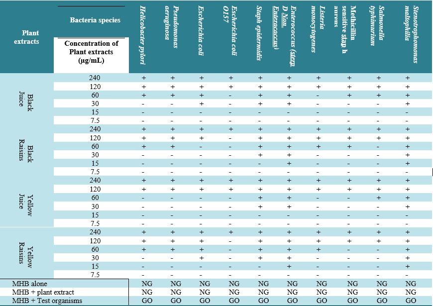
Note: Negative control (MHB alone or MHB + plant extract), NG: No growth
Positive control (MHB + test organisms), GO: Growth observed
MHB: Mueller Hinton Broth
Table 3. The result of MBC assay
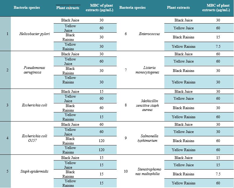
Table 4. The Pseudomonas aeruginosa biofilm inhibition presented for each extract corresponding to different concentrations
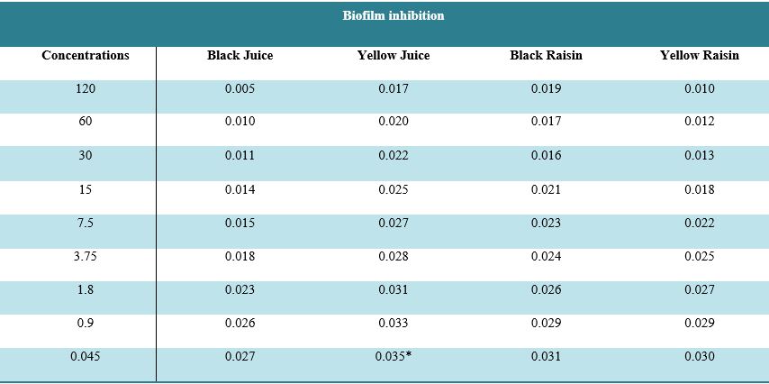
*Represents the maximum inhibition in biofilm growth for Pseudomonas aeruginosa.
Control: LB Broth+Pseudomonas aeruginosa.
3.5 Wound Healing activity
After evaluating the cytotoxic and antimicrobial activities of the extracts, a scratch assay was carried out on fibroblast cells. None of the extracts at a concentration of 30 µg/mL had no effect on increasing cell proliferation. At lower concentrations of the black and yellow grape juice, an increase in wound-healing activity was observed. Interestingly, the black and yellow raisin extracts did not promote cell proliferation and even appeared to inhibit it; therefore, no scratch closure was observed. Nevertheless, no cell death was observed after seven days. Yellow juice extract, at concentrations of 7.5 and 15 µg/mL, showed greater wound-healing activity than black grape juice and the control on fibroblast cells. It should be noted that scratch closure by yellow juice, black juice, and control samples was evaluated after 12, 16, and 18 hours, respectively. Furthermore, Fibroblast cells exhibited increased proliferation following treatment with these extracts.
4. Discussion
The antibiotic resistance represents an existential threat to the continued control of infections. Unfortunately, it has increased due to overpopulation, the increased use of antibiotics in clinics, and poor sewage disposal systems, among other factors (23).
To overcome the emergence of AMR, various researchers have focused on the antimicrobial activity of plant extracts against different bacteria and resistant strains (24).
Grape (Vitis spp.) is one of the most widely grown fruit crops in the world. Its products, such as grape juice, jams, and raisins, are also essential commodities in the global market.
Grape pomace, as the richest source of PCs among fruits, is a potential source of natural antioxidant and antimicrobial agents effective against various bacterial strains (25, 26).
Gram-negative bacteria have an outer membrane of the cell wall made up of structural lipopolysaccharides, so it is impermeable to lipophilic solutions, unlike Gram-positive bacteria, which do not have this outer membrane (27). However, Cueva et al. reported that Gram-negative bacteria were more susceptible than Gram-positive bacteria to grape extracts, suggesting that the antimicrobial activity of grape-derived phytochemicals may depend on their chemical composition and the bacterial species tested rather than solely on the Gram-staining characteristics (28). The study performed by Filocamo et al. also showed that among the Gram-negative bacteria, E. coli was the only susceptible strain (MIC and MBC of 2000 μg/mL) to grape juice (29). Silván et al. demonstrated that the grape seed extract had a strong capacity to inhibit Campylobacter spp growth (30). In contrast, another study revealed that S. Typhimurium and E. coli were resistant at all concentrations of grape polyphenols (31). Additionally, a study by Dias et al. found that Gram-positive intestinal pathogens (L. monocytogenes, S. aureus, and Enterococcus faecalis) are more sensitive to extracts of grape stems than Gram-negative pathogens (P. aeruginosa, E. coli, and Klebsiella pneumoniae) (32). A recent study shows that using recommended amounts of grape waste extracts can significantly reduce the presence of S. aureus and E. coli bacterial strains (33).
In this study, the MIC and MBC findings demonstrated that the antibacterial activity of the grape-derived extracts was species-dependent. Lower MIC values observed for Enterococcus and S. maltophilia, particularly with the raisin extracts, indicate a higher susceptibility of these organisms to the bioactive compounds present in the extracts. In contrast, E. coli O157 exhibited the highest MIC values. Among the tested extracts, black raisin and yellow grape juice exhibited the greatest overall inhibitory activity against the tested bacterial isolates. Among the bacterial strains, P. aeruginosa and Enterococcus were the most susceptible to all extracts. Based on Figure 2, similarities between the average effects of different extract concentrations on bacteria, including E.coli, S. Typhimurium, methicillin-sensitive S. aureus, and H. pylori, can be observed. Additionally, the maximum range of the average growth of the inhibition zone was observed for Enterococcus (~30 mm) in yellow raisins, and the minimum was for E. coli O157 (~3 mm) in yellow juice. In this research, the results of the two-way ANOVA showed, in most cases, no direct link between increasing the rate of each extract concentration and the zone of inhibition produced by the different bacterial strains investigated. In Figure 4, yellow raisins and yellow juice extracts displayed a linear relationship between increased concentration and inhibition zone diameter for Enterococcus; however, for the other two extracts, a direct connection was not observed. Methicillin-sensitive S. aureus showed no explicit suppression among the increased concentration and inhibition zone. In most countries, nosocomial infections (NIs) are a significant medical problem (34). P. aeruginosa was the fourth most common nosocomial infection, following E. coli, K. pneumoniae, and S. aureus, accounting for 7.96% (35). P. aeruginosa is an opportunistic pathogen that infects patients with cystic fibrosis, burns wounds, immunodeficiency, chronic obstructive pulmonary disorder (COPD), cancer, and severe infection requiring ventilation, such as coronavirus disease 2019 (COVID-19) (35). Eradication of P. aeruginosa has become increasingly complex due to its remarkable capacity to resist antibiotics (36). Phenolic compounds, such as those found in grape extracts, are considered critical natural molecules due to their ability to damage bacterial cell walls and influence biofilm formation in different bacterial strains (37, 38). These results showed that black grape juice reduces biofilm formation, and in addition to black grape juice, extracts from yellow raisins can also effectively prevent biofilm structures formed by bacterial strains. These findings are consistent with other reports on antimicrobial herbal drugs. For example, Thymus vulgaris and Origanum vulgare extracts inhibit bacterial growth by damaging cell walls and interfering with biofilm formation (39). Similarly, Curcuma longa (turmeric) and Allium sativum (garlic) show broad-spectrum antibacterial effects, comparable to the activity observed with grape and raisin extracts (40). The healing of skin wounds, particularly chronic wounds, remains a clinical emergency (22). Grape seed extract displayed remarkable wound-healing activity via accelerated wound closure rate, enhancing Transforming growth factor β1 (TGF-β1), vascular endothelial growth factor (VEGF), collagen expression, and suppressing inflammatory markers such as Tumor necrosis factor α (TNF-α) and Interleukin-1β (IL-1β) (41-43). Hemmati et al. showed that proanthocyanidins, a class of polyphenols, in grape seed (GS) oil trigger the release of VEGF and could be an effective wound healing agent (44). It is interesting to know that the antioxidant potential of proanthocyanidins from grape seeds is 20 times greater than that of vitamin C and 50 times greater than that of vitamin E (45). The study demonstrated that the yellow and black juice extracts were effective in increasing the proliferation of fibroblast cells. Surprisingly, black and yellow raisins did not affect cell proliferation, and even cell growth was suspended. As mentioned before, after seven days of testing, no cell death was observed. The cause of this phenomenon is obscure. These results, together with our results, indicate the positive role of grape extracts in promoting wound healing. Collectively, our results underscore the potential of grape and raisin extracts, predominantly yellow raisins and black grape juice, as natural agents with antimicrobial and wound healing properties. However, variability in efficacy across extract types and target bacterial strains highlights the need for further phytochemical analysis and in vivo validation. One limitation of the present study is that all experiments were performed in vitro; thus, the observed antimicrobial and wound healing effects may not fully translate to in vivo conditions. Additionally, only a limited number of bacterial and fungal strains were examined, and only selected concentrations of grape and raisin extracts were tested. Further studies are required to investigate dose-dependent effects, different extraction methods, and in vivo efficacy to validate these findings.
5. Conclusion
Grape and raisin extracts, particularly those from yellow raisins and black grapes, exhibited notable antibacterial activities against different bacterial strains and demonstrated wound healing properties with minimal cytotoxicity. These findings support their potential as natural therapeutic agents for combating bacterial infections and promoting tissue repair.
6. Declarations
6.1 Acknowledgments
We are grateful to Ali Hematian and Mojtaba Maleki for their time and expert knowledge.
6.2 Ethical Considerations
This study did not involve human participants or animals. Therefore, ethical approval was not required.
6.3 Authors' Contributions
S.Aghaei: methodology, writing (original draft), sample collection; K. Heidarzadi: conceptualization; S. Shokri: writing, editing; R. Amini: sample collection, methodology; I. Pakzad, A. Keshavarzi and M. Taherikalani: conceptualization; Sh. Mahmoudvand: review and editing; N. Ansari: methodology; Z. Ramezannia: review and editing; Gh.Kalvandi and F.Azizi Jalilian: materials provision, project administration, methodology; Final approval: All authors.
6.4 Conflict of Interest
The authors declare that they have no conflict of interest.
6.5 Fund or Financial Support
This research was funded by Ilam University of Medical Sciences, Ilam, Iran, grant number 918054/151.
6.6 Using Artificial Intelligence Tools (AI Tools)
The authors did not use AI tools.
Background & Objective: This study examines the in vitro antimicrobial properties of black and yellow grape juice and raisin extracts against 12 Gram-positive and Gram-negative bacterial strains, as well as five Candida species.
Materials & Methods: Cell viability was assessed by MTT assay, antibacterial activity by disk diffusion, MIC and MBC tests, biofilm inhibition by a microtiter plate method, and wound healing potential by a scratch assay.
Results: MTT assay showed that black and yellow grape juices and yellow raisins were non-toxic up to 30 µg/mL, while black raisins were safe only up to 15 µg/mL. Disk diffusion demonstrated antibacterial activity of all extracts against most tested bacterial except A. baumannii and Methicillin-resistant S. aureus. The strongest inhibition zones were observed for P. aeruginosa, Enterococcus, S. epidermidis, L. monocytogenes, and S. maltophilia, varying by extract type, while E. coli and E. coli O157 showed the lowest sensitivity. None of the extracts showed antifungal activity against Candida species. The lowest MIC observed for the yellow and black raisin extracts was 15 µg/mL against Enterococcus and S. maltophilia. In the yellow and black grape juice, the lowest MIC against S. epidermidis, Enterococcus, and S. maltophilia was 30 µg/mL. Black grape juice showed the greatest inhibition of P. aeruginosa biofilm formation. Both yellow and black grape juices promoted wound healing.
Conclusion: Grape extracts may serve as promising natural sources for the development of novel antibacterial agents against the tested Gram-positive and Gram-negative bacteria.
Keywords: Antibacterial activity, Biofilm inhibition, Grape extracts, Natural compounds, Wound healing
1. Introduction
Today, in many parts of the developing world, between 70% and 95% of people continue to rely on traditional medicine (TM), and many countries have integrated herbal medicine into their mainstream healthcare systems through regulations (8). Several studies have investigated the effectiveness of plant extracts and secondary metabolites that are endowed with antimicrobial properties, either alone or in combination with existing drugs (9-12). The low cost, biocompatibility, and effectiveness of herbal drugs make them an attractive source for the development of antimicrobial agents that enhance the biological activity of existing drugs (13, 14). Examples of medicinal plants with broad-spectrum antimicrobial activities include Allium sativum (garlic), Azadirachta indica (neem), Curcuma longa (15).
Grapes (Vitis vinifera L.) and their derivatives, including juice, pomace, and raisins, are rich sources of polyphenols such as proanthocyanidins, flavonoids, and resveratrol. These compounds exhibit antioxidant, anti-inflammatory, anticancer, antibacterial, and antifungal activities. Previous studies have shown that grape polyphenols can interact with bacterial cell walls, alter membrane permeability, inhibit microbial enzymes, and reduce biofilm formation (16). Therefore, evaluating the antimicrobial effects of grape and raisin extracts provides insights into their potential as natural alternatives or adjunctive therapies for treating infectious diseases. Furthermore, in addition to their antimicrobial activity, grape extracts have been reported to promote wound healing by enhancing fibroblast proliferation, stimulating angiogenesis, and modulating inflammatory responses. This dual functionality makes grape and raisin extracts promising candidates for applications in both infection control and tissue repair (17).
This study aimed to evaluate the effects of black and yellow grape juices, as well as raisin extracts, on wound healing and their antibacterial and antifungal activities. Additionally, the cytotoxicity of these extracts in fibroblast cells and their effects at safe concentrations on standard bacterial strains, clinical isolates, biofilm formation, and wound healing were also assessed.
2. Materials and Methods
2.1 Extracts
Black and yellow raisins from Ilam Gardens were purchased from a local grocery store. Subsequently, the raisins were shade-dried and then then dried in a hot-air oven. Fifty grams of dried powdered raisins were added in 320 mL of hydroalcoholic solution (70/30) and then shaken (140 rpm, room temperature) for 72 hours. Afterwards, the extract was filtered through Whatman No. 1 filter paper (S&S, Germany).
Subsequently, the solvent was removed using a rotary evaporator and the remaining extract was dried in forced-air drying oven for 48 hours. The concentrated extract was preserved in a sterile 50 mL Falcon tube at 4°C.
Fresh black and yellow grapes were manually crushed and filtered, and aliquots of the juice were stored at -20°C. Before use, juice samples were thawed and filtered through a 0.22 μm membrane filter.
The extracts were adjusted to pH 7.4 and diluted in the culture medium to achieve the desired concentrations for all cell culture experiments.
This standardized procedure ensured reproducibility and suitability for in vitro assays.
2.2 MTT assay
Vero cells, a fibroblast-like cell line, were used for the cytotoxicity assay. These cells were obtained from the National Cell Bank of Iran (Pasteur Institute, Tehran, Iran).
The cells were cultured in Dulbecco's Modified Eagle's Medium (DMEM) supplemented with 10% fetal calf serum (FCS) (Gibco, USA), penicillin, and streptomycin (60 μg/mL).
Cells were incubated at 37°C with 5% carbon dioxide (CO₂).
After 24–48 hours, the cells formed a monolayer and were subsequently passage into 96-well plates. Then, nine concentrations of the extracts, 60, 30, 15, 7.5, 3.75, 1.8, 0.93, 0.46, and 0.23 µg/mL, were added to the culture medium. The cytotoxicity was evaluated after 24, 48, and 72 hours using the colorimetric MTT (3-(4,5-dimethylthiazol-2-yl)-2,5-diphenyltetrazolium bromide) assay (18).
2.3 Bacterial strains
Gram-positive bacterial strains including Methicillin-resistant Staphylococcus aureus (ATCC700698), Methicillin-sensitive Staphylococcus aureus (ATCC 29247), Listeria monocytogenes (ATCC 35152), Group D non-enterococcal streptococcus (ATCC 33317), Staphylococcus epidermidis (ATCC 14990) and Gram-negative bacterial strains: Pseudomonas aeruginosa (ATCC 15442) , Stenotrophomonas maltophilia ( ATCC 13637), Acinetobacter baumannii (ATCC 17978), Salmonella Typhimurium (ATCC 13311), Escherichia coli (ATCC 25922), Escherichia coli O157 (ATCC 43894) and clinical isolates of Helicobacter pylori isolated from biopsies at the department of Medical Microbiology, Ilam University of Medical Sciences, were included to the survey. In addition, five fungal species were included in the study: Candida glabrata (ATCC 90030), Candida parapsilosis (ATCC 7330), Candida albicans (ATCC 14053), Candida tropicalis (ATCC MYA-3404), and Candida krusei (ATCC 6258).
2.4 Isolation of H. pylori
A homogenized suspension of antral biopsy specimens in sterile normal saline was prepared for the isolation of H. pylori. This suspension was then inoculated onto Brucella medium (MIRMEDIA, Iran) plates with 7% sheep blood, supplemented with the antibiotics including Vancomycin (10 mg/L), Trimethoprim (5 mg/L), and Polymyxin B (2.5 IU) to inhibit the growth of contaminating bacteria, under microaerophilic conditions using Anaerocult C (Merck, Germany) at 37°C. The plates were incubated for 48 hours; subsequently, oxidase, urease, and catalase tests, as well as Giemsa and Gram staining, confirmed the identification of the bacteria as H. pylori (19).
2.5 Bacterial strains and growth conditions
Stock cultures of the available bacterial strains in the microbiology collection were used to prepare 24-hour cultures on Mueller-Hinton agar (MIRMEDIA, Iran). Cultures were then incubated at 37°C. Due to the specific growth requirements of H. pylori, Brucella agar (MIRMEDIA, Iran) supplemented with sheep blood and appropriate antibiotics, along with GasPak Type C, was used to generate microaerophilic conditions. After 48 hours of incubation, and upon confirming adequate bacterial growth and the absence of transition to the coccoid form, the bacteria were prepared for subsequent experiments.
Disks for the disk diffusion assay (Padtan Teb, Iran) were prepared from 7.5, 15, and 30 µg/mL of black and yellow grape juices and raisins. Suspensions of all microorganisms were prepared to a turbidity equivalent to the 0.5 McFarland standard. Then, all bacterial strains were cultured on Mueller-Hinton agar, and H. pylori was cultured in Brucella agar with 7% sheep blood. For each bacterial strain, three disks containing the different grape extracts, along with one blank disk serving as the negative control, were placed on each agar plate. Control disks containing the appropriate reference antimicrobial agents were included for each bacterial strain and fungal species to compare the inhibition zones produced by the extracts with those of the corresponding standard antimicrobial agents .Tetracycline was used for H. pylori, Erythromycin for A. baumannii, Oxamycin for S. epidermidis, Methicillin-sensitive and methicillin-resistant S. aureus, Gentamicin for L. monocytogenes, Enterococcus, P. aeruginosa, Chloramphenicol for E. coli, E. coli O157, S. Typhimurium, S. maltophilia and finally for all five fungi amphotericin B. After 24 hours of incubation for the bacterial and fungal strains and 48 hours for H. pylori, the diameters of the inhibition zones for control disks and disks with different concentrations of extracts were measured (20). Results were evaluated and analyzed using two-way ANOVA, and the susceptibility of the bacterial and fungal strains and fungal species to extract disks was reported.
2.6 Minimum inhibitory concentration (MIC) and minimum bactericidal concentration (MBC)
The antimicrobial activity of juice and raisin extracts was examined against bacterial strains that were susceptible to the extracts in the disk diffusion test, using the broth macrodilution method to determine the MIC and MBC, as recommended by the according to the Clinical and Laboratory Standards Institute (CLSI) guidelines (20).
2.7 Biofilm
To study the inhibitory effect of the extracts on P. aeruginosa biofilm formation, a microtiter plate-based method was employed. In this method, 96-well flat-bottom microplates (SPL, Korea) were used. An 18-hour culture of P. aeruginosa culture was used to prepare a microbial suspension in Luria-Bertani (LB) broth (MIRMEDIA, Iran). The turbidity of the suspension was adjusted to an absorbance (OD600) of 0.4–0.6 by measuring its absorbance via a spectrophotometer at 600 nm. A total volume of 190 μL, consisting of the extract and LB broth, was dispensed into each well, followed by the addition of 10 μL of the bacterial suspension. The final concentrations of the extracts in the wells were 120, 60, 30, 15, 7.5, 3.75, 1.8, 0.93, and 0.45 mg/mL. Each extract dilution was tested in duplicate. LB broth was used as a negative control. The microplate was incubated at 37°C with shaking at 30 rpm for 18-20 hours. After incubation, the contents of the wells were gently discarded and rinsed with distilled water. The wells were stained with 0.1% crystal violet (Merck, Germany) for 10 minutes at room temperature and then washed three times with distilled water. In the final stage, 200 μL of 95% ethanol (Merck, Germany) was added to each well. The absorbance was measured at 492 nm using an ELISA reader (21).
2.8 Wound Healing activity
Three final concentrations of the extracts (7.5, 15, and 30 µg/mL) were prepared in DMEM. Fibroblast cells were cultured in a six-well plate (SPL, Korea) at a density of 5 × 10⁴ cells per well. The monolayer was gently scratched with a sterile 1 mL pipette tip. After scratching, the medium was removed, and the wells were washed twice with PBS. Fresh medium containing 5% FBS (Gibco, USA) and corresponding treatments was added to each well. Plates were incubated at 37°C with 5% CO₂. After 6, 8, 12, 14, 16, 18, 24, 36, and 48 hours of incubation, scratch closure was evaluated. In this test, the width of the scratches on the plate was measured and considered as a positive control and untreated scratched cells represented the control (22).
3. Result
3.1 MTT assay
The results of the cytotoxicity test are presented in Table 1. The cytotoxicity dose-response in 24 to 72 hours indicated that black and yellow juice, as well as yellow raisins, share the same non-toxic dose at 30 µg/mL, followed by black raisins at 15 µg/mL. Cell viability showed no significant difference between black and yellow juice at non-toxic doses (P > 0.05).
3.2 Disk diffusion
The grape juice and raisin extracts were tested against Gram-positive and Gram-negative bacterial strains, as well as five fungi, for their antimicrobial activities.
Table 1: Non-toxic concentrations of grape and raisin extracts in MTT assay
2.1 Extracts
Black and yellow raisins from Ilam Gardens were purchased from a local grocery store. Subsequently, the raisins were shade-dried and then then dried in a hot-air oven. Fifty grams of dried powdered raisins were added in 320 mL of hydroalcoholic solution (70/30) and then shaken (140 rpm, room temperature) for 72 hours. Afterwards, the extract was filtered through Whatman No. 1 filter paper (S&S, Germany).
Subsequently, the solvent was removed using a rotary evaporator and the remaining extract was dried in forced-air drying oven for 48 hours. The concentrated extract was preserved in a sterile 50 mL Falcon tube at 4°C.
Fresh black and yellow grapes were manually crushed and filtered, and aliquots of the juice were stored at -20°C. Before use, juice samples were thawed and filtered through a 0.22 μm membrane filter.
The extracts were adjusted to pH 7.4 and diluted in the culture medium to achieve the desired concentrations for all cell culture experiments.
This standardized procedure ensured reproducibility and suitability for in vitro assays.
2.2 MTT assay
Vero cells, a fibroblast-like cell line, were used for the cytotoxicity assay. These cells were obtained from the National Cell Bank of Iran (Pasteur Institute, Tehran, Iran).
The cells were cultured in Dulbecco's Modified Eagle's Medium (DMEM) supplemented with 10% fetal calf serum (FCS) (Gibco, USA), penicillin, and streptomycin (60 μg/mL).
Cells were incubated at 37°C with 5% carbon dioxide (CO₂).
After 24–48 hours, the cells formed a monolayer and were subsequently passage into 96-well plates. Then, nine concentrations of the extracts, 60, 30, 15, 7.5, 3.75, 1.8, 0.93, 0.46, and 0.23 µg/mL, were added to the culture medium. The cytotoxicity was evaluated after 24, 48, and 72 hours using the colorimetric MTT (3-(4,5-dimethylthiazol-2-yl)-2,5-diphenyltetrazolium bromide) assay (18).
2.3 Bacterial strains
Gram-positive bacterial strains including Methicillin-resistant Staphylococcus aureus (ATCC700698), Methicillin-sensitive Staphylococcus aureus (ATCC 29247), Listeria monocytogenes (ATCC 35152), Group D non-enterococcal streptococcus (ATCC 33317), Staphylococcus epidermidis (ATCC 14990) and Gram-negative bacterial strains: Pseudomonas aeruginosa (ATCC 15442) , Stenotrophomonas maltophilia ( ATCC 13637), Acinetobacter baumannii (ATCC 17978), Salmonella Typhimurium (ATCC 13311), Escherichia coli (ATCC 25922), Escherichia coli O157 (ATCC 43894) and clinical isolates of Helicobacter pylori isolated from biopsies at the department of Medical Microbiology, Ilam University of Medical Sciences, were included to the survey. In addition, five fungal species were included in the study: Candida glabrata (ATCC 90030), Candida parapsilosis (ATCC 7330), Candida albicans (ATCC 14053), Candida tropicalis (ATCC MYA-3404), and Candida krusei (ATCC 6258).
2.4 Isolation of H. pylori
A homogenized suspension of antral biopsy specimens in sterile normal saline was prepared for the isolation of H. pylori. This suspension was then inoculated onto Brucella medium (MIRMEDIA, Iran) plates with 7% sheep blood, supplemented with the antibiotics including Vancomycin (10 mg/L), Trimethoprim (5 mg/L), and Polymyxin B (2.5 IU) to inhibit the growth of contaminating bacteria, under microaerophilic conditions using Anaerocult C (Merck, Germany) at 37°C. The plates were incubated for 48 hours; subsequently, oxidase, urease, and catalase tests, as well as Giemsa and Gram staining, confirmed the identification of the bacteria as H. pylori (19).
2.5 Bacterial strains and growth conditions
Stock cultures of the available bacterial strains in the microbiology collection were used to prepare 24-hour cultures on Mueller-Hinton agar (MIRMEDIA, Iran). Cultures were then incubated at 37°C. Due to the specific growth requirements of H. pylori, Brucella agar (MIRMEDIA, Iran) supplemented with sheep blood and appropriate antibiotics, along with GasPak Type C, was used to generate microaerophilic conditions. After 48 hours of incubation, and upon confirming adequate bacterial growth and the absence of transition to the coccoid form, the bacteria were prepared for subsequent experiments.
Disks for the disk diffusion assay (Padtan Teb, Iran) were prepared from 7.5, 15, and 30 µg/mL of black and yellow grape juices and raisins. Suspensions of all microorganisms were prepared to a turbidity equivalent to the 0.5 McFarland standard. Then, all bacterial strains were cultured on Mueller-Hinton agar, and H. pylori was cultured in Brucella agar with 7% sheep blood. For each bacterial strain, three disks containing the different grape extracts, along with one blank disk serving as the negative control, were placed on each agar plate. Control disks containing the appropriate reference antimicrobial agents were included for each bacterial strain and fungal species to compare the inhibition zones produced by the extracts with those of the corresponding standard antimicrobial agents .Tetracycline was used for H. pylori, Erythromycin for A. baumannii, Oxamycin for S. epidermidis, Methicillin-sensitive and methicillin-resistant S. aureus, Gentamicin for L. monocytogenes, Enterococcus, P. aeruginosa, Chloramphenicol for E. coli, E. coli O157, S. Typhimurium, S. maltophilia and finally for all five fungi amphotericin B. After 24 hours of incubation for the bacterial and fungal strains and 48 hours for H. pylori, the diameters of the inhibition zones for control disks and disks with different concentrations of extracts were measured (20). Results were evaluated and analyzed using two-way ANOVA, and the susceptibility of the bacterial and fungal strains and fungal species to extract disks was reported.
2.6 Minimum inhibitory concentration (MIC) and minimum bactericidal concentration (MBC)
The antimicrobial activity of juice and raisin extracts was examined against bacterial strains that were susceptible to the extracts in the disk diffusion test, using the broth macrodilution method to determine the MIC and MBC, as recommended by the according to the Clinical and Laboratory Standards Institute (CLSI) guidelines (20).
2.7 Biofilm
To study the inhibitory effect of the extracts on P. aeruginosa biofilm formation, a microtiter plate-based method was employed. In this method, 96-well flat-bottom microplates (SPL, Korea) were used. An 18-hour culture of P. aeruginosa culture was used to prepare a microbial suspension in Luria-Bertani (LB) broth (MIRMEDIA, Iran). The turbidity of the suspension was adjusted to an absorbance (OD600) of 0.4–0.6 by measuring its absorbance via a spectrophotometer at 600 nm. A total volume of 190 μL, consisting of the extract and LB broth, was dispensed into each well, followed by the addition of 10 μL of the bacterial suspension. The final concentrations of the extracts in the wells were 120, 60, 30, 15, 7.5, 3.75, 1.8, 0.93, and 0.45 mg/mL. Each extract dilution was tested in duplicate. LB broth was used as a negative control. The microplate was incubated at 37°C with shaking at 30 rpm for 18-20 hours. After incubation, the contents of the wells were gently discarded and rinsed with distilled water. The wells were stained with 0.1% crystal violet (Merck, Germany) for 10 minutes at room temperature and then washed three times with distilled water. In the final stage, 200 μL of 95% ethanol (Merck, Germany) was added to each well. The absorbance was measured at 492 nm using an ELISA reader (21).
2.8 Wound Healing activity
Three final concentrations of the extracts (7.5, 15, and 30 µg/mL) were prepared in DMEM. Fibroblast cells were cultured in a six-well plate (SPL, Korea) at a density of 5 × 10⁴ cells per well. The monolayer was gently scratched with a sterile 1 mL pipette tip. After scratching, the medium was removed, and the wells were washed twice with PBS. Fresh medium containing 5% FBS (Gibco, USA) and corresponding treatments was added to each well. Plates were incubated at 37°C with 5% CO₂. After 6, 8, 12, 14, 16, 18, 24, 36, and 48 hours of incubation, scratch closure was evaluated. In this test, the width of the scratches on the plate was measured and considered as a positive control and untreated scratched cells represented the control (22).
3. Result
3.1 MTT assay
The results of the cytotoxicity test are presented in Table 1. The cytotoxicity dose-response in 24 to 72 hours indicated that black and yellow juice, as well as yellow raisins, share the same non-toxic dose at 30 µg/mL, followed by black raisins at 15 µg/mL. Cell viability showed no significant difference between black and yellow juice at non-toxic doses (P > 0.05).
3.2 Disk diffusion
The grape juice and raisin extracts were tested against Gram-positive and Gram-negative bacterial strains, as well as five fungi, for their antimicrobial activities.
Table 1: Non-toxic concentrations of grape and raisin extracts in MTT assay
Disk diffusion tests were performed for all four types of extracts at concentrations of 7.5, 15, and 30 µg/mL. All bacteria, except A. baumannii and Methicillin-resistant S. aureus, showed zones of inhibition. The maximum diameter growth of inhibition zone in millimeters (mm) for black raisin extracts in the concentration of 30 µg/mL belonged to (P. aeruginosa~25 mm), (L. monocytogenes~24 mm), (Enterococcus~22 mm), (S. maltophilia~21 mm), (S. epidermidis~20 mm), for yellow raisin extracts maximum growth was observed in (Enterococcus~30mm), (P. aeruginosa~28 mm), (S. epidermidis~24 mm), (L. monocytogenes~23 mm), regarding the yellow juice the maximum rate was visible in (P. aeruginosa and Enterococcus~29 mm), (L. monocytogenes~25 mm), (S. maltophilia~23 mm), and finally for black grape extract the maximum rate was dominant in (P. aeruginosa~26 mm) and (Enterococcus and S. epidermidis~20 mm).
The minimum growth rate, as indicated by the inhibition zone diameter, was observed for all four extracts at different concentrations in E. coli and E. coli O157. At a concentration of 7.5 µg/mL, the three extracts (yellow raisin, black raisin, and yellow raisin juice) showed no inhibitory effect against E. coli O157. The highest average sensitivity among the bacterial strains was observed in Enterococcus toward the yellow raisin extracts, and the lowest sensitivity belonged to E. coli O157 in yellow grape juice (Figure 1). As shown in Figure 2, among the tested extracts, black raisin and yellow grape juice exhibited the highest overall inhibitory effects. Among the bacterial strains, P. aeruginosa and Enterococcus were the most susceptible to all extracts. The zone of inhibition produced by two bacterial strains, S. maltophilia, and E. coli, are shown in Figure 3.
The results of Figure 4 show the relationship between increased concentration and the growth inhibition zone.
The anti-fungal activity of the extracts against C.glabrata,
C.parapsilosis, C.albicans, C. tropicalis, and C.krusei was not detected; therefore, the tests were continued only with the sensitive bacterial strains.
3.3 MIC and MBC
The MIC values of all the plant extracts against the microorganisms are shown in Table 2. The concentrations of the plant extracts considered for the MIC assay were 7.5, 15, 30, 60, 120, and 240 µg/mL. These extracts exhibited antibacterial activity against the tested microorganisms, although the degree of activity varied among the bacterial species. The lowest MIC (highest antibacterial activity) for yellow and black raisin extracts was 15 µg/mL against Enterococcus, and S. maltophilia. In the yellow and black grape juice, the lowest MIC for S. epidermidis, S. maltophilia, and Enterococcus was observed at a concentration of 30 µg/mL. The highest MIC value (240 µg/mL), indicating the lowest antibacterial activity, was observed for both yellow and black raisin extracts against E. coli O157.
Results from the MBC assay supported the data obtained from the disk diffusion assay and MIC assay (Table 3).
Figure 1. Growth rate of inhibition zone diameter. Bacteria name: 1. Helicobacter pylori, 2. Pseudomonas aeruginosa, 3. Escherichia coli, 4. Escherichia coli O157, 5. Staph epidermidis, 6. Enterococcus, 7. Listeria monocytogenes, 8. Methicillin sensitive staph aureus, 9. Salmonella typhimurium, 10. Stenotrophomonas maltophilia. (Prepared by Authors, 2026).
Figure 2. Estimated marginal means of inhibition zone diameters for 10 bacterial strains exposed to four grape-derived extracts (Black Raisins, Yellow Raisins, Black Juice, and Yellow Juice. (Prepared by Authors, 2026).
Figure 3. The diameter of inhibition zone in Stenotrophomonas maltophilia and E.coli. Left box bacteria: Stenotrophomonas maltophilia, Right box bacteria: Escherichia coli. First row in each box from left to right: Black raisins, raisins; Second row in each box from left to right: Black juice, yellow juice. (Prepared by Authors, 2026).
Figure 4. Relationship between increased concentration and growth inhibition zone. (Prepared by Authors, 2026).
Table 2. Minimum inhibitory concentrations of the plant extracts against the pathogenic organisms
Note: Negative control (MHB alone or MHB + plant extract), NG: No growth
Positive control (MHB + test organisms), GO: Growth observed
MHB: Mueller Hinton Broth
Table 3. The result of MBC assay
3.4 Inhibitory effect on biofilm
The inhibitory effects of extracts were evaluated for P.aeruginosa (Table 4).
At a concentration of 120 µg/mL (which is higher than the concentrations obtained from the MBC assay), black juice displayed the foremost activity in suppressing biofilm formation at an OD of 0.005. However, cytotoxic concentrations of the other two extracts, including yellow juice and black raisin extracts, were not significantly effective in inhibiting the biofilm formation. In this test, P. aeruginosa in LB broth was used as the control, which showed in 0.014 OD. At concentrations of 30 µg/mL and above, black juice showed the lowest OD, followed by yellow raisin extract, with an OD below 0.013. The lowest OD was at 0.005 in 120 µg/mL, indicating the maximum biofilm inhibition for black juice.
The inhibitory effects of extracts were evaluated for P.aeruginosa (Table 4).
At a concentration of 120 µg/mL (which is higher than the concentrations obtained from the MBC assay), black juice displayed the foremost activity in suppressing biofilm formation at an OD of 0.005. However, cytotoxic concentrations of the other two extracts, including yellow juice and black raisin extracts, were not significantly effective in inhibiting the biofilm formation. In this test, P. aeruginosa in LB broth was used as the control, which showed in 0.014 OD. At concentrations of 30 µg/mL and above, black juice showed the lowest OD, followed by yellow raisin extract, with an OD below 0.013. The lowest OD was at 0.005 in 120 µg/mL, indicating the maximum biofilm inhibition for black juice.
Table 4. The Pseudomonas aeruginosa biofilm inhibition presented for each extract corresponding to different concentrations
*Represents the maximum inhibition in biofilm growth for Pseudomonas aeruginosa.
Control: LB Broth+Pseudomonas aeruginosa.
3.5 Wound Healing activity
After evaluating the cytotoxic and antimicrobial activities of the extracts, a scratch assay was carried out on fibroblast cells. None of the extracts at a concentration of 30 µg/mL had no effect on increasing cell proliferation. At lower concentrations of the black and yellow grape juice, an increase in wound-healing activity was observed. Interestingly, the black and yellow raisin extracts did not promote cell proliferation and even appeared to inhibit it; therefore, no scratch closure was observed. Nevertheless, no cell death was observed after seven days. Yellow juice extract, at concentrations of 7.5 and 15 µg/mL, showed greater wound-healing activity than black grape juice and the control on fibroblast cells. It should be noted that scratch closure by yellow juice, black juice, and control samples was evaluated after 12, 16, and 18 hours, respectively. Furthermore, Fibroblast cells exhibited increased proliferation following treatment with these extracts.
4. Discussion
The antibiotic resistance represents an existential threat to the continued control of infections. Unfortunately, it has increased due to overpopulation, the increased use of antibiotics in clinics, and poor sewage disposal systems, among other factors (23).
To overcome the emergence of AMR, various researchers have focused on the antimicrobial activity of plant extracts against different bacteria and resistant strains (24).
Grape (Vitis spp.) is one of the most widely grown fruit crops in the world. Its products, such as grape juice, jams, and raisins, are also essential commodities in the global market.
Grape pomace, as the richest source of PCs among fruits, is a potential source of natural antioxidant and antimicrobial agents effective against various bacterial strains (25, 26).
Gram-negative bacteria have an outer membrane of the cell wall made up of structural lipopolysaccharides, so it is impermeable to lipophilic solutions, unlike Gram-positive bacteria, which do not have this outer membrane (27). However, Cueva et al. reported that Gram-negative bacteria were more susceptible than Gram-positive bacteria to grape extracts, suggesting that the antimicrobial activity of grape-derived phytochemicals may depend on their chemical composition and the bacterial species tested rather than solely on the Gram-staining characteristics (28). The study performed by Filocamo et al. also showed that among the Gram-negative bacteria, E. coli was the only susceptible strain (MIC and MBC of 2000 μg/mL) to grape juice (29). Silván et al. demonstrated that the grape seed extract had a strong capacity to inhibit Campylobacter spp growth (30). In contrast, another study revealed that S. Typhimurium and E. coli were resistant at all concentrations of grape polyphenols (31). Additionally, a study by Dias et al. found that Gram-positive intestinal pathogens (L. monocytogenes, S. aureus, and Enterococcus faecalis) are more sensitive to extracts of grape stems than Gram-negative pathogens (P. aeruginosa, E. coli, and Klebsiella pneumoniae) (32). A recent study shows that using recommended amounts of grape waste extracts can significantly reduce the presence of S. aureus and E. coli bacterial strains (33).
In this study, the MIC and MBC findings demonstrated that the antibacterial activity of the grape-derived extracts was species-dependent. Lower MIC values observed for Enterococcus and S. maltophilia, particularly with the raisin extracts, indicate a higher susceptibility of these organisms to the bioactive compounds present in the extracts. In contrast, E. coli O157 exhibited the highest MIC values. Among the tested extracts, black raisin and yellow grape juice exhibited the greatest overall inhibitory activity against the tested bacterial isolates. Among the bacterial strains, P. aeruginosa and Enterococcus were the most susceptible to all extracts. Based on Figure 2, similarities between the average effects of different extract concentrations on bacteria, including E.coli, S. Typhimurium, methicillin-sensitive S. aureus, and H. pylori, can be observed. Additionally, the maximum range of the average growth of the inhibition zone was observed for Enterococcus (~30 mm) in yellow raisins, and the minimum was for E. coli O157 (~3 mm) in yellow juice. In this research, the results of the two-way ANOVA showed, in most cases, no direct link between increasing the rate of each extract concentration and the zone of inhibition produced by the different bacterial strains investigated. In Figure 4, yellow raisins and yellow juice extracts displayed a linear relationship between increased concentration and inhibition zone diameter for Enterococcus; however, for the other two extracts, a direct connection was not observed. Methicillin-sensitive S. aureus showed no explicit suppression among the increased concentration and inhibition zone. In most countries, nosocomial infections (NIs) are a significant medical problem (34). P. aeruginosa was the fourth most common nosocomial infection, following E. coli, K. pneumoniae, and S. aureus, accounting for 7.96% (35). P. aeruginosa is an opportunistic pathogen that infects patients with cystic fibrosis, burns wounds, immunodeficiency, chronic obstructive pulmonary disorder (COPD), cancer, and severe infection requiring ventilation, such as coronavirus disease 2019 (COVID-19) (35). Eradication of P. aeruginosa has become increasingly complex due to its remarkable capacity to resist antibiotics (36). Phenolic compounds, such as those found in grape extracts, are considered critical natural molecules due to their ability to damage bacterial cell walls and influence biofilm formation in different bacterial strains (37, 38). These results showed that black grape juice reduces biofilm formation, and in addition to black grape juice, extracts from yellow raisins can also effectively prevent biofilm structures formed by bacterial strains. These findings are consistent with other reports on antimicrobial herbal drugs. For example, Thymus vulgaris and Origanum vulgare extracts inhibit bacterial growth by damaging cell walls and interfering with biofilm formation (39). Similarly, Curcuma longa (turmeric) and Allium sativum (garlic) show broad-spectrum antibacterial effects, comparable to the activity observed with grape and raisin extracts (40). The healing of skin wounds, particularly chronic wounds, remains a clinical emergency (22). Grape seed extract displayed remarkable wound-healing activity via accelerated wound closure rate, enhancing Transforming growth factor β1 (TGF-β1), vascular endothelial growth factor (VEGF), collagen expression, and suppressing inflammatory markers such as Tumor necrosis factor α (TNF-α) and Interleukin-1β (IL-1β) (41-43). Hemmati et al. showed that proanthocyanidins, a class of polyphenols, in grape seed (GS) oil trigger the release of VEGF and could be an effective wound healing agent (44). It is interesting to know that the antioxidant potential of proanthocyanidins from grape seeds is 20 times greater than that of vitamin C and 50 times greater than that of vitamin E (45). The study demonstrated that the yellow and black juice extracts were effective in increasing the proliferation of fibroblast cells. Surprisingly, black and yellow raisins did not affect cell proliferation, and even cell growth was suspended. As mentioned before, after seven days of testing, no cell death was observed. The cause of this phenomenon is obscure. These results, together with our results, indicate the positive role of grape extracts in promoting wound healing. Collectively, our results underscore the potential of grape and raisin extracts, predominantly yellow raisins and black grape juice, as natural agents with antimicrobial and wound healing properties. However, variability in efficacy across extract types and target bacterial strains highlights the need for further phytochemical analysis and in vivo validation. One limitation of the present study is that all experiments were performed in vitro; thus, the observed antimicrobial and wound healing effects may not fully translate to in vivo conditions. Additionally, only a limited number of bacterial and fungal strains were examined, and only selected concentrations of grape and raisin extracts were tested. Further studies are required to investigate dose-dependent effects, different extraction methods, and in vivo efficacy to validate these findings.
5. Conclusion
Grape and raisin extracts, particularly those from yellow raisins and black grapes, exhibited notable antibacterial activities against different bacterial strains and demonstrated wound healing properties with minimal cytotoxicity. These findings support their potential as natural therapeutic agents for combating bacterial infections and promoting tissue repair.
6. Declarations
6.1 Acknowledgments
We are grateful to Ali Hematian and Mojtaba Maleki for their time and expert knowledge.
6.2 Ethical Considerations
This study did not involve human participants or animals. Therefore, ethical approval was not required.
6.3 Authors' Contributions
S.Aghaei: methodology, writing (original draft), sample collection; K. Heidarzadi: conceptualization; S. Shokri: writing, editing; R. Amini: sample collection, methodology; I. Pakzad, A. Keshavarzi and M. Taherikalani: conceptualization; Sh. Mahmoudvand: review and editing; N. Ansari: methodology; Z. Ramezannia: review and editing; Gh.Kalvandi and F.Azizi Jalilian: materials provision, project administration, methodology; Final approval: All authors.
6.4 Conflict of Interest
The authors declare that they have no conflict of interest.
6.5 Fund or Financial Support
This research was funded by Ilam University of Medical Sciences, Ilam, Iran, grant number 918054/151.
6.6 Using Artificial Intelligence Tools (AI Tools)
The authors did not use AI tools.
Type of Study: Original Research Article |
Subject:
Medical Biology
Received: 2025/10/3 | Accepted: 2026/03/17 | Published: 2026/05/20
Received: 2025/10/3 | Accepted: 2026/03/17 | Published: 2026/05/20
References
1. Murugaiyan J, Kumar PA, Rao GS, Iskandar K, Hawser S, Hays JP, et al. Progress in alternative strategies to combat antimicrobial resistance: Focus on antibiotics. Antibiotics. 2022;11(2):200. [DOI:10.3390/antibiotics11020200] [PMID] [PMCID]
2. Maleki MH, Sekawi Z, Soroush S, Azizi-Jalilian F, Asadollahi K, Mohammadi S, et al. Phenotypic and genotypic characteristics of tetracycline resistant Acinetobacter baumannii isolates from nosocomial infections at Tehran hospitals. Iran J Basic Med Sci. 2014;17(1):21-6.
3. Hasanvand A, Ghafourian S, Taherikalani M, Jalilian FA, Sadeghifard N, Pakzad I. Antiseptic resistance in methicillin sensitive and methicillin resistant staphylococcus aureus iIsolates from some major hospitals, Iran. Recent Pat Antiinfect Drug Discov. 2015;10(2):105-12. [DOI:10.2174/1574891X10666150623093259] [PMID]
4. Chassagne F, Samarakoon T, Porras G, Lyles JT, Dettweiler M, Marquez L, et al. A systematic review of plants with antibacterial activities: A taxonomic and phylogenetic perspective. Front Pharmacol. 2020;11:586548. [DOI:10.3389/fphar.2020.586548] [PMID] [PMCID]
5. Yang X, Ye W, Qi Y, Ying Y, Xia Z. Overcoming multidrug resistance in bacteria through antibiotics delivery in surface-engineered nano-cargos: recent developments for future nano-antibiotics. Front Bioeng Biotechnol. 2021;9:696514. [DOI:10.3389/fbioe.2021.696514] [PMID] [PMCID]
6. Gajdács M, Urbán E, Stájer A, Baráth Z. Antimicrobial resistance in the context of the sustainable development goals: A brief review. Eur J Investig Health Psychol Educ. 2021;11(1):71-82. [DOI:10.3390/ejihpe11010006] [PMID] [PMCID]
7. Chouhan S, Sharma K, Guleria S. Antimicrobial activity of some essential oils-present status and future perspectives. Medicines (Basel). 2017;4(3):58. [DOI:10.3390/medicines4030058] [PMID] [PMCID]
8. Ekor M. The growing use of herbal medicines: issues relating to adverse reactions and challenges in monitoring safety. Front Pharmacol. 2014;4:177. [DOI:10.3389/fphar.2013.00177] [PMID] [PMCID]
9. Saavedra MJ, Borges A, Dias C, Aires A, Bennett RN, Rosa ES, et al. Antimicrobial activity of phenolics and glucosinolate hydrolysis products and their synergy with streptomycin against pathogenic bacteria. Med Chem. 2010;6(3):174-83. [DOI:10.2174/1573406411006030174] [PMID]
10. Djeussi DE, Noumedem JA, Seukep JA, Fankam AG, Voukeng IK, Tankeo SB, et al. Antibacterial activities of selected edible plants extracts against multidrug-resistant Gram-negative bacteria. BMC Complement Altern Med. 2013;13:164. [DOI:10.1186/1472-6882-13-164] [PMID] [PMCID]
11. Abreu AC, Serra SC, Borges A, Saavedra MJ, Salgado AJ, Simões M. Evaluation of the best method to assess antibiotic potentiation by phytochemicals against Staphylococcus aureus. Diagn Microbiol Infect Dis. 2014;79(2):125-34. [DOI:10.1016/j.diagmicrobio.2014.03.002] [PMID]
12. Monte J, Abreu AC, Borges A, Simões LC, Simões M. Antimicrobial activity of selected phytochemicals against Escherichia coli and Staphylococcus aureus and their biofilms. Pathogens. 2014;3(2):473-98. [DOI:10.3390/pathogens3020473] [PMID] [PMCID]
13. Parthasarathy A, Borrego EJ, Savka MA, Dobson RCJ, Hudson AO. Amino acid-derived defense metabolites from plants: A potential source to facilitate novel antimicrobial development. J Biol Chem. 2021;296:100438. [DOI:10.1016/j.jbc.2021.100438] [PMID] [PMCID]
14. Ali Roozegar M, Azizi Jalilian F, Reza Havasian M, Panahi J, Pakzad I. Antimicrobial effect of Pistacia atlantica leaf extract. Bioinformation. 2016;12(1):19-21. [DOI:10.6026/97320630012019] [PMID] [PMCID]
15. El-Saadony MT, Saad AM, Mohammed DM, Korma SA, Alshahrani MY, Ahmed AE, et al. Medicinal plants: bioactive compounds, biological activities, combating multidrug-resistant microorganisms, and human health benefits - a comprehensive review. Front Immunol. 2025;16:1491777. [DOI:10.3389/fimmu.2025.1491777] [PMID] [PMCID]
16. Gerardi C, Pinto L, Baruzzi F, Giovinazzo G. Comparison of antibacterial and antioxidant properties of red (cv. Negramaro) and white (cv. Fiano) skin pomace extracts. Molecules. 2021;26(19).
17. Ajit A, Vishnu AG, Varkey P. Incorporation of grape seed extract towards wound care product development. 3 Biotech. 2021;11(6):261. [DOI:10.1007/s13205-021-02826-4] [PMID] [PMCID]
18. Frattaruolo L, Carullo G, Brindisi M, Mazzotta S, Bellissimo L, Rago V, et al. Antioxidant and anti-inflammatory activities of flavanones from Glycyrrhiza glabra L. (licorice) leaf phytocomplexes: identification of licoflavanone as a modulator of NF-kB/MAPK pathway. Antioxidants (Basel, Switzerland). 2019;8(6). [DOI:10.3390/antiox8060186] [PMID] [PMCID]
19. Choi KD, Kim JM, Baik GH, Park JC, Jung HK, Ryu HS, et al. Helicobacter pylori isolation and antibiotic susceptibility testing using rapidly frozen biopsy samples. Korean J Helicobacter Up Gastrointest Res. 2024;24(4):360-4. [DOI:10.7704/kjhugr.2024.0063] [PMID] [PMCID]
20. Hemeg HA, Moussa IM, Ibrahim S, Dawoud TM, Alhaji JH, Mubarak AS, et al. Antimicrobial effect of different herbal plant extracts against different microbial population. Saudi J Biol Sci. 2020;27(12):3221-7. [DOI:10.1016/j.sjbs.2020.08.015] [PMID] [PMCID]
21. Stepanovic S, Vukovic D, Hola V, Bonaventura GD, Djukic S, Ćirkovic I, et al. Quantification of biofilm in microtiter plates: overview of testing conditions and practical recommendations for assessment of biofilm production by staphylococci. APMIS. 2007;115(8):891-9. [DOI:10.1111/j.1600-0463.2007.apm_630.x] [PMID]
22. Carullo G, Sciubba F, Governa P, Mazzotta S, Frattaruolo L, Grillo G, et al. Mantonico and pecorello grape seed extracts: Chemical characterization and evaluation of in vitro wound-healing and anti-inflammatory activities. Pharmaceuticals (Basel, Switzerland). 2020;13(5):97. [DOI:10.3390/ph13050097] [PMID] [PMCID]
23. Aslam B, Wang W, Arshad MI, Khurshid M, Muzammil S, Rasool MH, et al. Antibiotic resistance: a rundown of a global crisis. Infect Drug Resist. 2018;11:1645-58. [DOI:10.2147/IDR.S173867] [PMID] [PMCID]
24. Badet C. Chapter 65 - Antibacterial activity of grape (Vitis vinifera, Vitis rotundifolia) seeds. In: Preedy VR, Watson RR, Patel VB, editors. Nuts and Seeds in Health and Disease Prevention. San Diego: Academic Press; 2011. p. 545-52. [DOI:10.1016/B978-0-12-375688-6.10065-9]
25. Xu Y, Burton S, Kim C, Sismour E. Phenolic compounds, antioxidant, and antibacterial properties of pomace extracts from four Virginia-grown grape varieties. Food Sci Nutr. 2016;4(1):125-33. [DOI:10.1002/fsn3.264] [PMID] [PMCID]
26. Cosme F, Pinto T, Vilela A. Phenolic compounds and antioxidant activity in grape juices: A chemical and sensory view. Beverages. 2018;4(1):22. [DOI:10.3390/beverages4010022]
27. Bertani B, Ruiz N. Function and biogenesis of lipopolysaccharides. EcoSal Plus. 2018;8(1). [DOI:10.1128/ecosalplus.esp-0001-2018] [PMID] [PMCID]
28. Cueva C, Mingo S, Muñoz-González I, Bustos I, Requena T, del Campo R, et al. Antibacterial activity of wine phenolic compounds and oenological extracts against potential respiratory pathogens. Lett Appl Microbiol. 2012;54(6):557-63. [DOI:10.1111/j.1472-765X.2012.03248.x] [PMID]
29. Filocamo A, Bisignano C, Mandalari G, Navarra M. In vitro antimicrobial activity and effect on biofilm production of a white grape juice (Vitis vinifera) extract. Evid Base Complement Alternat Med .2015;2015:856243. [DOI:10.1155/2015/856243] [PMID] [PMCID]
30. Silván JM, Mingo E, Hidalgo M, de Pascual-Teresa S, Carrascosa AV, Martinez-Rodriguez AJ. Antibacterial activity of a grape seed extract and its fractions against Campylobacter spp. Food Control. 2013;29(1):25-31. [DOI:10.1016/j.foodcont.2012.05.063]
31. Yadav D, Kumar A, Kumar P, Mishra D. Antimicrobial properties of black grape (Vitis vinifera L.) peel extracts against antibiotic-resistant pathogenic bacteria and toxin producing molds. Indian J Pharmacol. 2015;47(6):663-7. [DOI:10.4103/0253-7613.169591] [PMID] [PMCID]
32. Dias C, Domínguez-Perles R, Aires A, Teixeira A, Rosa E, Barros A, et al. Phytochemistry and activity against digestive pathogens of grape (Vitis vinifera L.) stem's (poly) phenolic extracts.Food Sci Technol. 2015;61(1):25-32. [DOI:10.1016/j.lwt.2014.11.033]
33. Dakheli MJ. Effects of grape and pomegranate waste extracts on poultry carcasses microbial, chemical, and sensory attributes in slaughterhouse. Food Sci Nutr. 2020;8(10):5622-30. [DOI:10.1002/fsn3.1840] [PMID] [PMCID]
34. Pezhman B, Fatemeh R, Amir R, Mahboobeh R, Mohammad F. Nosocomial infections in an Iranian educational hospital: an evaluation study of the Iranian nosocomial infection surveillance system. BMC Infect Dis. 2021;21(1):1256. [DOI:10.1186/s12879-021-06948-1] [PMID] [PMCID]
35. Qin S, Xiao W, Zhou C, Pu Q, Deng X, Lan L, et al. Pseudomonas aeruginosa: pathogenesis, virulence factors, antibiotic resistance, interaction with host, technology advances and emerging therapeutics. Sig Transduct Target Ther. 2022;7(1):199. [DOI:10.1038/s41392-022-01056-1] [PMID] [PMCID]
36. Pang Z, Raudonis R, Glick BR, Lin TJ, Cheng Z. Antibiotic resistance in Pseudomonas aeruginosa: mechanisms and alternative therapeutic strategies. Biotechnol Adv. 2019;37(1):177-92. [DOI:10.1016/j.biotechadv.2018.11.013] [PMID] [PMCID]
37. Hassan YI, Kosir V, Yin X, Ross K, Diarra MS. Grape pomace as a promising antimicrobial alternative in feed: A Critical Review. J Agric Food Chem. 2019;67(35):9705-18. [DOI:10.1021/acs.jafc.9b02861] [PMID]
38. Efenberger-Szmechtyk M, Nowak A, Czyzowska A. Plant extracts rich in polyphenols: antibacterial agents and natural preservatives for meat and meat products. Crit Rev Food Sci Nutr. 2021;61(1):149-78. [DOI:10.1080/10408398.2020.1722060] [PMID]
39. Carezzano ME, Sotelo JP, Primo E, Reinoso EB, Paletti Rovey MF, Demo MS, et al. Inhibitory effect of Thymus vulgaris and Origanum vulgare essential oils on virulence factors of phytopathogenic Pseudomonas syringae strains. Plant Biology. 2017;19(4):599-607. [DOI:10.1111/plb.12572] [PMID]
40. Wada NM, Ambi AA, Ibrahim AA, Bello SK, Umar A, James DT. Antimicrobial activity of extracts of Turmeric (Curcuma longa) and Garlic (Aliium sativum) against selected bacterial clinical isolates. Medit J Infect Microb Antimicrob. 2021.
41. Khanna S, Venojarvi M, Roy S, Sharma N, Trikha P, Bagchi D, et al. Dermal wound healing properties of redox-active grape seed proanthocyanidins. Free Radic Biol Med. 2002;33(8):1089-96. [DOI:10.1016/S0891-5849(02)00999-1] [PMID]
42. Khanna S, Roy S, Bagchi D, Bagchi M, Sen CK. Upregulation of oxidant-induced VEGF expression in cultured keratinocytes by a grape seed proanthocyanidin extract. Free Radic Biol Med. 2001;31(1):38-42. [DOI:10.1016/S0891-5849(01)00544-5] [PMID]
43. Al-Warhi T, Zahran EM, Selim S, Al-Sanea MM, Ghoneim MM, Maher SA, et al. Antioxidant and wound healing potential of Vitis vinifera seeds supported by phytochemical characterization and docking studies. Antioxidants (Basel, Switzerland). 2022;11(5). [DOI:10.3390/antiox11050881] [PMID] [PMCID]
44. Hemmati AA, Foroozan M, Houshmand G, Moosavi ZB, Bahadoram M, Maram NS. The topical effect of grape seed extract 2% cream on surgery wound healing. Glob J Health Sci. 2014;7(3):52-8. [DOI:10.5539/gjhs.v7n3p52] [PMID] [PMCID]
45. Shi J, Yu J, Pohorly JE, Kakuda Y. Polyphenolics in grape seeds-biochemistry and functionality. J Med Food. 2003;6(4):291-9. [DOI:10.1089/109662003772519831] [PMID]
| Rights and permissions | |
 |
This work is licensed under a Creative Commons Attribution-NonCommercial 4.0 International License. |
Copyright Policy
Journal of Advances in Medical and Biomedical Research by Zanjan University of Medical Sciences is licensed under CC BY-NC 4.0