
Fri, Nov 13, 2026
[Archive]
Volume 34, Issue 4 (July & August 2026)
J Adv Med Biomed Res 2026, 34(4): 331-337 |
Back to browse issues page


Ethics code: IR.IAU.Z.REC.1396,101


Download citation:
BibTeX | RIS | EndNote | Medlars | ProCite | Reference Manager | RefWorks
Send citation to:


BibTeX | RIS | EndNote | Medlars | ProCite | Reference Manager | RefWorks
Send citation to:
Shahmohamadi F, Sadeghi Z, Davoudi Z. Assessment of Serum Expression of lncRNA MHENCR, miR-425, and PI3K in Iranian Patients with Melanoma. J Adv Med Biomed Res 2026; 34 (4) :331-337
URL: http://journal.zums.ac.ir/article-1-7898-en.html
URL: http://journal.zums.ac.ir/article-1-7898-en.html
Assessment of Serum Expression of lncRNA MHENCR, miR-425, and PI3K in Iranian Patients with Melanoma


1- National center for health insurance research, Tehran, Iran
2- Cellular and Molecular Research Center, Research Institute of Cellular and Molecular Sciences in Infectious Diseases, Zahedan University of Medical Sciences, Zahedan, Iran
3- Department of Medical Biotechnology, Zanjan University of Medical Science, Zanjan, Iran ,davoudibiotech@gmail.com
2- Cellular and Molecular Research Center, Research Institute of Cellular and Molecular Sciences in Infectious Diseases, Zahedan University of Medical Sciences, Zahedan, Iran
3- Department of Medical Biotechnology, Zanjan University of Medical Science, Zanjan, Iran ,
Full-Text [PDF 336 kb]
(93 Downloads)
| Abstract (HTML) (754 Views)
1. Introduction
Table 1. Clinical Characteristics of Melanoma Patients.
Table 2. Sequences of Primers.
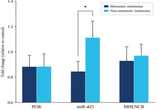
Figure 1. The expression levels of PI3K, miR-425, and MHENC in patients with metastatic melanoma and non-metastatic melanoma. (Prepared by Authors, 2026).
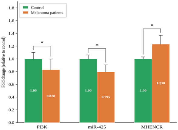
Figure 2. The expression levels of PI3K, miR-425, and MHENC in patients with metastatic and non-metastatic melanoma compared to the control group. (Prepared by Authors, 2026).
Full-Text: (48 Views)
1. Introduction
Melanoma ranks among the most aggressive human cancers. It develops from the uncontrolled proliferation of melanocytes and most frequently linked to UV radiation exposure, which causes genetic mutations and promotes tumor formation. Annually, approximately 320,000 new cases of cutaneous melanoma are diagnosed worldwide, resulting in about 57,000 deaths. In Iran, it accounts for 5.2% of all malignancies. Unlike squamous and basal cell cancers, melanomas can spread unpredictably and potentially affect any organ in the body. Moreover, melanoma demonstrates significant resistance to current therapies, especially during its metastatic stage. There are three main subtypes of melanoma: Superficial Spreading Melanoma (SSM), which comprises approximately 70% of cases and is linked to UV exposure or arises from naevi; Lentigo Maligna Melanoma (LMM), responsible for 4 to 15% of cases and associated with UV exposure and older age; and Nodular Melanoma (NMM), making up 5% of cases and characterized by rapid progression and a high rate of metastasis. Other variants are uncommon (1-5).
MicroRNAs (miRNAs) are a class of small noncoding RNAs (containing 21 to 23 nucleotides) that regulate gene expression by interacting with the 3′-UTR of specific mRNA targets, resulting in either degradation of the transcript or inhibition of translation. MiRNAs are derived from coding and non-coding transcriptional units found in introns, exons, or intergenic regions. Their generation involves a complex, multistep process that requires the coordinated action of proteins in both the nucleus and cytoplasm (6-8). MiRNAs are involved in oncogenic processes through their ability to regulate the expression of various tumor suppressor genes and oncogenes, as well as by triggering pathways associated with cancer progression. Over the past few years, profiling and sequencing analyses have provided evidence that miRNA signatures can be utilized in cancer for classification, diagnostic purposes, and prognosis assessment. Researchers have evaluated the diverse patterns of miRNA expression and their functions across various types of cancer. MiRNAs may function either as oncogenes or tumor suppressors depending on the cancerous context. Oncogenic miRNAs enhance tumor development by inhibiting tumor suppressor genes or apoptosis-related genes, while tumor suppressor miRNAs suppress cancer progression by targeting oncogenes or genes that regulate cell cycle progression and promote apoptosis (9-11).
Melanoma miRNA profiles revealed that various miRNAs modulate gene translation or stability depending on the disease stage, highlighting their crucial role in regulating processes linked to melanoma progression. Recent findings have revealed that certain types of miRNAs, such as miR-98, miR-200c, miRNA-106b, miR-33a, and miR-524-5p, are implicated in melanoma progression. Considering previous research, it indicates that miR-425 may perform multiple roles by modulating various downstream targets across different types of cancer. This microRNA functions as a tumor suppressor by targeting IGF1 in melanoma (12-14).
Another type of functional noncoding RNA is long noncoding RNA (lncRNA), which is longer than 200 nucleotides and is involved in the regulation of many tumor suppressor and oncogene expressions (15,16). Their role has been reported in various tumors, including melanoma.
MHENCR (melanoma highly expressed noncoding RNA), as a noncoding RNA, plays a crucial role in tumorigenesis. Studies have shown that it is upregulated in melanoma and promotes melanoma progression. This lncRNA specifically binds to miR-425 and miR-489, leading to increased expression of their target genes, namely IGF-1 and SPIN1, thereby activating the PI3K-Akt signaling pathway.
This pathway plays a crucial role in the coordinated regulation of metabolism, as well as cell growth and proliferation. In many types of cancer, components of this pathway become dysregulated, ultimately, resulting in the inhibition of apoptosis and an increase in cell proliferation and growth (17-21).
Therefore, given that investigating the MHENCR/miR‑425/PI3K axis in melanoma may offer new insights into the molecular mechanisms of tumorigenesis and metastasis, this study examined the expression of these genes and evaluated how alterations in their expression correlate with patients’ clinical characteristics compared to healthy controls. Considering the growing burden of melanoma and limitations of tissue‑based diagnostics, there is also a particular need for non‑invasive biomarkers in Iranian populations to facilitate early detection, risk stratification, and disease monitoring through accessible and minimally invasive approaches.
2. Materials and Methods
2.1 Sample Collection
In this study, 34 samples were collected from Razi Specialized Dermatology Hospital in Tehran and Vali-e-Asr Specialized Hospital in Zanjan. These included 17 melanoma samples and 17 normal samples from healthy individuals. All samples were obtained with the patients’ informed consent and ethical approval from the ethics committee (IR.IAU.Z.REC.1396.101). Healthy individuals and patients were between 20 and 80 years old, and blood samples were collected in EDTA-containing tubes and stored at 20°C until use. Clinical characteristics of melanoma patients are shown in the Table 1.
2.2 RNA Extraction and cDNA Synthesis
RNA was extracted using the Roche RNA extraction kit following the manufacturer’s protocol. RNA quality was assessed using both NanoDrop spectrophotometry and agarose gel electrophoresis. cDNA was synthesized using the Takara PrimeScript RT reagent kit. During cDNA synthesis, appropriate volumes of buffer, easy dilution solution, oligo dT primers, random 6-mer primers, reverse transcriptase enzyme, total RNA, and master mix were combined and incubated in a PCR machine under the following temperature conditions: 37°C for 15 minutes, 85°C for 5 seconds, and 4°C for 5 minutes. Upon completion, the samples were stored at -20 °C. Stem-loop primers targeting mir33a and the control mir199 were employed during cDNA synthesis from miRNA. The sequences of these primers are listed in Table 2. Primers were designed and validated for specificity using Primer-BLAST (NCBI) and for secondary structure using OligoAnalyzer.
2.3 Quantitative Real-Time PCR (qRT-PCR)
The real-time PCR reaction was carried out in a total volume of 25 µl, containing 1 µl of forward primer, 1 µl of reverse primer, 12.5 µl of SYBR Green, 8.5 µl of water, and 2 µl of cDNA. The samples were then placed in the real-time PCR machine and run for 45 cycles with a total reaction volume of 25 µl, using the annealing temperatures specific to each primer, as indicated in Table 2. Primer sequences used in the study are listed in Table 2. The melting curve was analyzed at the end of each reaction. Experiments were performed in triplicate to ensure reproducibility and accuracy of the results (22).
2.4 Statistical Analysis
Statistical analysis was performed using SPSS software. Expression changes between tumor and normal samples were assessed using the 2-ΔΔCT method. The significance of the results was evaluated by the t-test, with p-values less than 0.05 considered statistically significant.
Table 1. Clinical Characteristics of Melanoma Patients.
| Patient ID | Age Year |
Sex | Diagnosis | Treatment | Pathology Report | Family History |
| 1 | 84 | Male | Melanoma of the ear | Cryotherapy Surgical excision |
R/O: Melanoma Skin biopsy (right ear) Ki67/HMB45 |
Negative |
| 2 | 54 | Female | Melanoma of the finger | Cryotherapy Excision of lesion |
R/O: Melanoma in situ Clark I-II C44/7 M8720/3 |
Negative |
| 3 | 53 | Male | Melanoma of the sole | Emergency surgery on sole and proximal 4th–5th fingers | Malignant melanoma Invasive vertical growth Ulceration: present Clark III-IV Perineural invasion: none C44/7 M8720/3 |
Negative |
| 4 | 60 | Male | Melanoma of the toe | Referred to Cancer Institute for surgery | Clark III IHC supports malignant melanoma Breslow: ~2.2 mm Nail unit biopsy C44.6 M8744.3 |
Negative |
| 5 | 60 | Female | Melanoma of the sole | Emergency referral for surgery | Clark III C44.6 MM70.3 |
Negative |
| 6 | 71 | Male | Melanoma of scalp and auricle | 2 sessions cryotherapy Biopsy of auricle Excision of lesion |
Lentigo maligna Thickness: 0.73 mm Clark I No ulceration 6 mm margin recommended |
Negative |
| 7 | 55 | Female | Melanoma of the right hand | Emergency referral for surgery | Clark I-II Melanocyte hyperplasia Nail biopsy (right hand, 3rd finger) Margins involved C44.6 M8721.3 |
Negative |
| 8 | 57 | Male | Melanoma of the scalp | Surgery with 2 cm margins Wound bed prep STSG repair |
Malignant melanoma IHC: C44.4 M8721.3 |
Negative |
| 9 | 64 | Male | Melanoma of the sole | Two surgeries Radiotherapy + Chemotherapy (DTIC, Cisplatin, Carboplatin, Ketril, Plasil, Dexamethasone) |
Malignant melanoma Sole lesion |
Negative |
| 10 | 61 | Male | Metastatic melanoma (lung & liver) | Surgery Radiotherapy Chemotherapy (Interferon, G-CSF, DTIC 400 mg, Carboplatin 600 mg, Cisplatin 300 mg, Ketril, Plasil, Dexamethasone) Port insertion |
Metastatic malignant melanoma Lung and liver involvement |
Positive (colon, thyroid, laryngeal cancer in relatives) |
| 11 | 74 | Female | Metastatic melanoma (lung & brain) | Surgery Chemotherapy (Alimta, Metformin, Atorvastatin, Metoprolol) |
Metastatic malignant melanoma Lung and brain involvement |
Negative |
| 12 | 75 | Female | Metastatic melanoma (brain & lung) | Surgery Radiotherapy Chemotherapy (IFN-α2, Dacarbazine, Docetaxel, Dexamethasone, Ketril, Plasil) |
Metastatic malignant melanoma Extensive brain and lung involvement |
Negative |
| 13 | 54 | Female | Anal melanoma | Surgery and removal of anal lesions Chemotherapy (DTIC, Cisplatin, Metformin 500 mg, Glibenclamide 5 mg, Ketril, Plasil, Ranitidine) |
Malignant melanoma Anal region |
Negative |
| 14 | 69 | Male | Foot melanoma with DVT & cellulitis | Two surgeries Chemotherapy (Interferon alpha, Enoxaparin, Vancomycin) |
Malignant melanoma Foot with DVT and cellulitis |
Negative |
| 15 | 26 | Female | Metastatic melanoma (liver) | Surgery Ascitic fluid tap Chemotherapy (DTIC, Domperidone, Ketril, Plasil, Ranitidine) PTC insertion to open bile duct |
Metastatic malignant melanoma Liver involvement with ascites |
Negative |
| 16 | 57 | Male | Nasal melanoma | Surgery and excision Chemotherapy + Port insertion (Interferon, DTIC 600, G-CSF 300, Cisplatin, Carboplatin, Imatinib 400) |
Malignant melanoma Nasal region |
Negative |
| 17 | 62 | Male | Melanoma (2nd finger) + axillary mass + pulmonary metastasis | Lung surgery + excision Chemotherapy (DTIC 375 mg, Carboplatin 25 mg, Taxol 110 mg) CT: multiple bilateral pulmonary metastatic nodules; hypodense liver lesion 38×36 mm (segment IV) |
Metastatic malignant melanoma Finger, axilla, lung and liver involvement |
Negative |
Table 2. Sequences of Primers.
| Annealing Temperature | Sequences | Name |
| 72 оC | 5'-GTT GGC TCT GGT GCA GGG TCC GAG GTA TTC GCA CCA GAG CCA ACG AAA GA-3' |
Stem loop mir-425 |
| 73 оC | 5'-GTT GGC TCT GGT GCA GGG TCC GAG GTA TTC GCA CCA GAG CCA ACA GGC AG -3' |
Stem loop mir-199 |
| 49 оC | 5'-ATG GTC AGT GGA CGG ACG-3' | MHENCR Forward |
| 47 оC | 5'-ACA GCA AGC ACA AGG GTG-3' | MHENCR Reverse |
| 54 оC | 5'-CTC TCC TGT GCT GGC TAC TGT-3' | PI3K Forward |
| 49 оC | 5'-GCT CTC GGT TGA TTC CAA ACT-3' | PI3K Reverse |
| 46 оC | 5'-TGT TTT TTG AAA GCG CTT TGG AA-3' | Mir-425 Forward |
| 48 оC | 5'-GTG CAG GGT CCG AGG T-3' | Mir-425 Reverse |
| 52 оC | 5'-GGG AAA TCG TGC GTG ACA TTA AG-3' | β-Actin Forward |
| 51 оC | 5'-TGT GTT GGC GTA CAG GTC TTT G -3' | β-Actin Reverse |
| 51 оC | 5'-ACC ACA GTC CAT GCC ATC AC-3' | GAPDH Forward |
| 50 оC | 5'-TCC ACC CTG TTG CTG TA -3' | GAPDH Reverse |
3. Result
3.1 Patient and Control Group Sample Characteristics
In this study, a total of 17 blood samples were collected from melanoma patients at the specialized dermatology hospitals Razi in Tehran and Hazrat Vali Asr in Zanjan. Additionally, 17 control samples were obtained from healthy individuals without melanoma. It is important to note that none of the individuals in the control group had melanoma, nor did they have any family history of the disease. Furthermore, the number of moles on the bodies of the healthy control individuals was fewer than 50. Informed consent was obtained from all participants, both patients and controls, prior to blood sample collection. Participants were free to withdraw from the study at any time.
3.2 Real-time PCR results
The expression levels of PI3K, mir425, and MHENC in the patient groups are presented in Figure 1 (p < 0.05). The expression levels of PI3K, mir425, and MHENC in patients with metastatic melanoma showed fold changes of 0.883, 0.845, and 0.93, respectively. In patients with non-metastatic melanoma, PI3K, mir425, and MHENC exhibited fold changes of 0.884, 1.112, and 0.97, respectively. When analyzing the overall expression across all patients relative to the control, PI3K and mir425 decreased by 0.828-fold and 0.795-fold, respectively (p < 0.05), whereas the MHENC expression increased by 1.23-fold (p < 0.05). The expression levels of PI3K, mir425, and MHENC compared to the control group are shown in Figure 2.
3.1 Patient and Control Group Sample Characteristics
In this study, a total of 17 blood samples were collected from melanoma patients at the specialized dermatology hospitals Razi in Tehran and Hazrat Vali Asr in Zanjan. Additionally, 17 control samples were obtained from healthy individuals without melanoma. It is important to note that none of the individuals in the control group had melanoma, nor did they have any family history of the disease. Furthermore, the number of moles on the bodies of the healthy control individuals was fewer than 50. Informed consent was obtained from all participants, both patients and controls, prior to blood sample collection. Participants were free to withdraw from the study at any time.
3.2 Real-time PCR results
The expression levels of PI3K, mir425, and MHENC in the patient groups are presented in Figure 1 (p < 0.05). The expression levels of PI3K, mir425, and MHENC in patients with metastatic melanoma showed fold changes of 0.883, 0.845, and 0.93, respectively. In patients with non-metastatic melanoma, PI3K, mir425, and MHENC exhibited fold changes of 0.884, 1.112, and 0.97, respectively. When analyzing the overall expression across all patients relative to the control, PI3K and mir425 decreased by 0.828-fold and 0.795-fold, respectively (p < 0.05), whereas the MHENC expression increased by 1.23-fold (p < 0.05). The expression levels of PI3K, mir425, and MHENC compared to the control group are shown in Figure 2.
Figure 1. The expression levels of PI3K, miR-425, and MHENC in patients with metastatic melanoma and non-metastatic melanoma. (Prepared by Authors, 2026).
Figure 2. The expression levels of PI3K, miR-425, and MHENC in patients with metastatic and non-metastatic melanoma compared to the control group. (Prepared by Authors, 2026).
4. Discussion
MicroRNA molecules play a crucial role at multiple stages of melanoma progression. These stages include the invasion of cancer cells into blood or lymphatic vessels, their survival within the circulatory or lymphatic systems, exit from these vessels, and the establishment of new tumor sites in distant organs.
Melanoma progression is closely linked to metabolic changes within cells, where miRNAs play a key regulatory role. Studies have demonstrated that miRNA expression is often dysregulated in various cancers, and specific miRNA expression profiles can classify tumor types and predict clinical outcomes in melanoma patients. This knowledge is particularly valuable because miRNA-targeted therapies have the potential to slow melanoma progression and enhance cancer cell sensitivity to treatments. Several miRNAs associated with melanoma have been identified that functionally interact with key signaling pathways, including NF-Κb, Wnt/β-catenin, MAPK/ERK, and PI3K/PTEN/Akt. Among these microRNAs are miR-9, miR-377, miR-21, miR-23b, miR-25, miR-425, miR-29, and others (23-26).
In 2017, Chen and colleagues investigated the role of a long noncoding RNA called MHENCR in melanoma progression. They found that miR-425/489 influences melanoma by regulating the PI3K-AKT pathway, which is active in many cancers. In this study, MHENCR was found to be highly expressed in melanoma tissues, especially in metastatic melanoma, and this increased expression was associated with shorter patient survival. Experiments showed that reducing MHENCR expression slowed melanoma cell growth, caused cell cycle arrest, and induced apoptosis. Additionally, the cells’ ability to spread to other parts of the body was reduced. MHENCR interacts with miR-425/489 target genes such as SPIN1 and IGF1, activating them. This indicates that MHENCR promotes melanoma growth by activating the PI3K-AKT pathway. Therefore, the interaction between miR-425/489 and MHENCR and the activation of the PI3K-AKT pathway may represent a new therapeutic target for melanoma. Mechanistically, MHENCR acts as a ceRNA by sponging miR-425 and miR-489, relieving their inhibitory effects on SPIN1 and IGF1. Upregulated IGF1 activates IGF1R, which recruits and activates PI3K, leading to the conversion of PIP2 to PIP3 and subsequent AKT phosphorylation. Activated AKT promotes cell survival and proliferation by inhibiting pro-apoptotic factors and activating mTORC1, while SPIN1 further amplifies this signaling cascade. Collectively, targeting this MHENCR/miR-425-489/PI3K-AKT axis may provide a novel and effective therapeutic strategy against melanoma (21). According to the findings of this study, we also observed in our study on melanoma patients that the expression level of MHENCR was increased in Iranian melanoma patients. In 2015, Liu P, et al. conducted a study on the role of miR-425. They reported that IGF-1 acts as a potential tumor suppressor in cancer and inhibits melanoma progression by regulating miR-425. Specifically, miR-425 functions as a tumor suppressor in melanoma (27). Consistent with Liu’s study, we found that miR-425 expression was decreased in melanoma patients compared to the control group. In another study, Shahbazi et al. investigated the anticancer properties of flavonoids by examining their effects on the PI3K/Akt signaling pathway. Dysregulation of this pathway triggers various cellular responses, including growth, proliferation, survival, and motility, all of which contribute to tumor progression. Flavonoids inhibit PI3K activity by initially activating ACT, which leads to suppression of the enzyme. It has been established that targeting the PI3K/Akt pathway can be effective in cancer prevention and treatment (28). Several studies have confirmed that dysregulation of lncRNAs and miRNAs through the PI3K/AKT signaling axis is a recurrent and clinically significant mechanism in melanoma pathogenesis, with AKT pathway activation reported in approximately 70% of melanoma cases. These findings collectively validate the biological relevance of the MHENCR/miR-425/PI3K-AKT network observed in our study and reinforce its potential as a therapeutic target in melanoma (29, 30). In this study, we also found that PI3K gene expression was reduced in melanoma patients compared to the control group, a decrease that may be attributed to treatments such as drug therapy and chemotherapy administered to these patients. Limitations of this study include the small sample size (n=17), lack of an independent validation cohort. Future studies with larger cohorts and independent validation will confirm these findings.
5. Conclusion
Our results showed that the expression level of lncRNA MHENCR was increased in melanoma samples, while the expression levels of miR-425 and PI3K were decreased. These changes may be influenced by drug and chemotherapy treatments. Considering the potential roles of these molecules in regulatory pathways related to melanoma, further studies are needed to thoroughly investigate their functions and mechanisms. This will provide evidence-based support for their potential use as diagnostic biomarkers or therapeutic targets in melanoma management.
6. Declarations
6.1 Acknowledgments
The authors would like to express their gratitude to the staff and pathology departments of Razi Hospital (Tehran) and Vali-e-Asr Hospital (Zanjan), as well as all patients and healthy volunteers who participated in this study.
6.2 Ethical Considerations
This study was conducted in accordance with the Declaration of Helsinki and approved by the Ethics Committee of Islamic Azad University, Zanjan Branch (Ethics Code: IR.IAU.Z.REC.1396.101). Written informed consent was obtained from all individual participants (both melanoma patients and healthy controls) prior to blood sample collection. Participants were informed of their right to withdraw from the study at any time without consequence.
6.3 Authors' Contributions
All authors contributed significantly to the conception, design, and execution of this study: Fatemeh Shahmohamadi: Conceptualization, sample collection and data curation, Zohre Sadeghi: Methodology, formal analysis, software/statistical analysis and writing – original draft preparation. Zahra Davoudi: Supervision, validation, and writing – review & editing. All authors read and approved the final manuscript.
6.4 Conflict of Interest
The authors declare that they have no known competing financial interests or personal relationships that could have appeared to influence the work reported in this paper.
6.5 Fund or Financial Support
This research received no specific grant from any funding agency in the public, commercial, or not-for-profit sectors.
6.6 Using Artificial Intelligence Tools (AI Tools)
The authors declare that no artificial intelligence (AI) tools or generative AI technologies were used in the writing, data analysis, or preparation of this manuscript.
MicroRNA molecules play a crucial role at multiple stages of melanoma progression. These stages include the invasion of cancer cells into blood or lymphatic vessels, their survival within the circulatory or lymphatic systems, exit from these vessels, and the establishment of new tumor sites in distant organs.
Melanoma progression is closely linked to metabolic changes within cells, where miRNAs play a key regulatory role. Studies have demonstrated that miRNA expression is often dysregulated in various cancers, and specific miRNA expression profiles can classify tumor types and predict clinical outcomes in melanoma patients. This knowledge is particularly valuable because miRNA-targeted therapies have the potential to slow melanoma progression and enhance cancer cell sensitivity to treatments. Several miRNAs associated with melanoma have been identified that functionally interact with key signaling pathways, including NF-Κb, Wnt/β-catenin, MAPK/ERK, and PI3K/PTEN/Akt. Among these microRNAs are miR-9, miR-377, miR-21, miR-23b, miR-25, miR-425, miR-29, and others (23-26).
In 2017, Chen and colleagues investigated the role of a long noncoding RNA called MHENCR in melanoma progression. They found that miR-425/489 influences melanoma by regulating the PI3K-AKT pathway, which is active in many cancers. In this study, MHENCR was found to be highly expressed in melanoma tissues, especially in metastatic melanoma, and this increased expression was associated with shorter patient survival. Experiments showed that reducing MHENCR expression slowed melanoma cell growth, caused cell cycle arrest, and induced apoptosis. Additionally, the cells’ ability to spread to other parts of the body was reduced. MHENCR interacts with miR-425/489 target genes such as SPIN1 and IGF1, activating them. This indicates that MHENCR promotes melanoma growth by activating the PI3K-AKT pathway. Therefore, the interaction between miR-425/489 and MHENCR and the activation of the PI3K-AKT pathway may represent a new therapeutic target for melanoma. Mechanistically, MHENCR acts as a ceRNA by sponging miR-425 and miR-489, relieving their inhibitory effects on SPIN1 and IGF1. Upregulated IGF1 activates IGF1R, which recruits and activates PI3K, leading to the conversion of PIP2 to PIP3 and subsequent AKT phosphorylation. Activated AKT promotes cell survival and proliferation by inhibiting pro-apoptotic factors and activating mTORC1, while SPIN1 further amplifies this signaling cascade. Collectively, targeting this MHENCR/miR-425-489/PI3K-AKT axis may provide a novel and effective therapeutic strategy against melanoma (21). According to the findings of this study, we also observed in our study on melanoma patients that the expression level of MHENCR was increased in Iranian melanoma patients. In 2015, Liu P, et al. conducted a study on the role of miR-425. They reported that IGF-1 acts as a potential tumor suppressor in cancer and inhibits melanoma progression by regulating miR-425. Specifically, miR-425 functions as a tumor suppressor in melanoma (27). Consistent with Liu’s study, we found that miR-425 expression was decreased in melanoma patients compared to the control group. In another study, Shahbazi et al. investigated the anticancer properties of flavonoids by examining their effects on the PI3K/Akt signaling pathway. Dysregulation of this pathway triggers various cellular responses, including growth, proliferation, survival, and motility, all of which contribute to tumor progression. Flavonoids inhibit PI3K activity by initially activating ACT, which leads to suppression of the enzyme. It has been established that targeting the PI3K/Akt pathway can be effective in cancer prevention and treatment (28). Several studies have confirmed that dysregulation of lncRNAs and miRNAs through the PI3K/AKT signaling axis is a recurrent and clinically significant mechanism in melanoma pathogenesis, with AKT pathway activation reported in approximately 70% of melanoma cases. These findings collectively validate the biological relevance of the MHENCR/miR-425/PI3K-AKT network observed in our study and reinforce its potential as a therapeutic target in melanoma (29, 30). In this study, we also found that PI3K gene expression was reduced in melanoma patients compared to the control group, a decrease that may be attributed to treatments such as drug therapy and chemotherapy administered to these patients. Limitations of this study include the small sample size (n=17), lack of an independent validation cohort. Future studies with larger cohorts and independent validation will confirm these findings.
5. Conclusion
Our results showed that the expression level of lncRNA MHENCR was increased in melanoma samples, while the expression levels of miR-425 and PI3K were decreased. These changes may be influenced by drug and chemotherapy treatments. Considering the potential roles of these molecules in regulatory pathways related to melanoma, further studies are needed to thoroughly investigate their functions and mechanisms. This will provide evidence-based support for their potential use as diagnostic biomarkers or therapeutic targets in melanoma management.
6. Declarations
6.1 Acknowledgments
The authors would like to express their gratitude to the staff and pathology departments of Razi Hospital (Tehran) and Vali-e-Asr Hospital (Zanjan), as well as all patients and healthy volunteers who participated in this study.
6.2 Ethical Considerations
This study was conducted in accordance with the Declaration of Helsinki and approved by the Ethics Committee of Islamic Azad University, Zanjan Branch (Ethics Code: IR.IAU.Z.REC.1396.101). Written informed consent was obtained from all individual participants (both melanoma patients and healthy controls) prior to blood sample collection. Participants were informed of their right to withdraw from the study at any time without consequence.
6.3 Authors' Contributions
All authors contributed significantly to the conception, design, and execution of this study: Fatemeh Shahmohamadi: Conceptualization, sample collection and data curation, Zohre Sadeghi: Methodology, formal analysis, software/statistical analysis and writing – original draft preparation. Zahra Davoudi: Supervision, validation, and writing – review & editing. All authors read and approved the final manuscript.
6.4 Conflict of Interest
The authors declare that they have no known competing financial interests or personal relationships that could have appeared to influence the work reported in this paper.
6.5 Fund or Financial Support
This research received no specific grant from any funding agency in the public, commercial, or not-for-profit sectors.
6.6 Using Artificial Intelligence Tools (AI Tools)
The authors declare that no artificial intelligence (AI) tools or generative AI technologies were used in the writing, data analysis, or preparation of this manuscript.
Type of Study: Original Research Article |
Subject:
Life Science
Received: 2026/04/25 | Accepted: 2026/06/22 | Published: 2026/08/4
Received: 2026/04/25 | Accepted: 2026/06/22 | Published: 2026/08/4
References
1. Liu C, Liu X, Hu L, Li X, Xin H, Zhu S. Global, regional, and national burden of cutaneous malignant melanoma from 1990 to 2021 and prediction to 2045. Front Oncol. 2024;14:1512942. [DOI:10.3389/fonc.2024.1512942] [PMID] [PMCID]
2. Roky AH, Islam MM, Ahasan AMF, Mostaq MS, Mahmud MZ, Amin MN, et al. Overview of skin cancer types and prevalence rates across continents. Cancer Pathog Ther. 2025;3(02):89-100. [DOI:10.1016/j.cpt.2024.08.002] [PMID] [PMCID]
3. Druskovich C, Kelley J, Aubrey J, Palladino L, Wright GP. A review of melanoma subtypes: genetic and treatment considerations. J Surg Oncol. 2025;131(3):356-64. [DOI:10.1002/jso.27953] [PMID] [PMCID]
4. Naik PP. Cutaneous malignant melanoma: a review of early diagnosis and management. World J Oncol. 2021;12(1):7. [DOI:10.14740/wjon1349] [PMID] [PMCID]
5. Rakhshan A, Moradi A, Masoudi E. Melanoma in Iranian childhood and adolescence: An Analysis of 14 Patients. Int J Cancer Manage. 2023;16(1). [DOI:10.5812/ijcm-121873]
6. He L, Hannon GJ. MicroRNAs: small RNAs with a big role in gene regulation. Nat Rev Genet. 2004;5(7):522-31. [DOI:10.1038/nrg1379] [PMID] []
7. Hudder A, Novak RF. MiRNAs: effectors of environmental influences on gene expression and disease. Toxicol Sci. 2008;103(2):228-40. [DOI:10.1093/toxsci/kfn033] [PMID] [PMCID]
8. Sevignani C, Calin GA, Siracusa LD, Croce CM. Mammalian microRNAs: a small world for fine-tuning gene expression. Mamm Genome. 2006;17(3):189-202. [DOI:10.1007/s00335-005-0066-3] [PMID] [PMCID]
9. Svoronos AA, Engelman DM, Slack FJ. OncomiR or tumor suppressor? The duplicity of microRNAs in cancer. Cancer Res. 2016;76(13):3666-70. [DOI:10.1158/0008-5472.CAN-16-0359] [PMID] [PMCID]
10. Otmani K, Lewalle P. Tumor suppressor miRNA in cancer cells and the tumor microenvironment: mechanism of deregulation and clinical implications. Front Oncol. 2021;11:708765. [DOI:10.3389/fonc.2021.708765] [PMID] [PMCID]
11. Shenouda SK, Alahari SK. MicroRNA function in cancer: oncogene or a tumor suppressor? Cancer Metastasis Rev. 2009;28(3):369-378. [DOI:10.1007/s10555-009-9188-5] [PMID]
12. Cruz ATD, Jasiulionis MG. miRNAs and melanoma: how are they connected? Dermatol Res Pract. 2012;2012:528345. [DOI:10.1155/2012/528345] [PMID] [PMCID]
13. Segura MF, Greenwald HS, Hanniford D, Osman I, Hernando E. MicroRNA and cutaneous melanoma: from discovery to prognosis and therapy. Carcinogenesis. 2012;33(10):1823-32. [DOI:10.1093/carcin/bgs205] [PMID] [PMCID]
14. Fattore L, Costantini S, Malpicci D, Ruggiero CF, Ascierto PA, Croce CM, et al. MicroRNAs in melanoma development and resistance to target therapy. Oncotarget. 2017;8(13):22262. [DOI:10.18632/oncotarget.14763] [PMID] [PMCID]
15. Mattick JS, Amaral PP, Carninci P, Carpenter S, Chang HY, Chen LL, et al. Long non-coding RNAs: definitions, functions, challenges and recommendations. Nat Rev Mol Cell Biol. 2023;24(6):430-47. [DOI:10.1038/s41580-022-00566-8] [PMID]
16. Nikoofar P, Azodian Ghajar H. Long non-coding RNAs: hidden regulators of cancer. Transl Res Urol. 2023;5(1):1-3.
17. Wozniak M, Czyz M. The functional role of long non-coding RNAs in melanoma. Cancers. 2021;13(19):4848. [DOI:10.3390/cancers13194848] [PMID] [PMCID]
18. Montico B, Giurato G, Pecoraro G, Salvati A, Covre A, Colizzi F, et al. The pleiotropic roles of circular and long noncoding RNAs in cutaneous melanoma. Mol Oncol. 2022;16(3):565-93. [DOI:10.1002/1878-0261.13034] [PMID] [PMCID]
19. Li P, Ma X, Gu X. The essential roles of lncRNAs/PI3K/AKT axis in gastrointestinal tumors. Front Cell Dev Biol. 2024;12:1442193. [DOI:10.3389/fcell.2024.1442193] [PMID] [PMCID]
20. Xiao Y, Xia Y, Wang Y, Xue C. Pathogenic roles of long noncoding RNAs in melanoma: implications in diagnosis and therapies. Genes Dis. 2023;10(1):113-25. [DOI:10.1016/j.gendis.2021.08.007] [PMID] [PMCID]
21. Chen X, Dong H, Liu S, Yu L, Yan D, Yao X, et al. Long noncoding RNA MHENCR promotes melanoma progression via regulating miR-425/489-mediated PI3K-Akt pathway. Am J Transl Res. 2017;9(1):90.
22. Zhu J, Deng J, Zhang L, Zhao J, Zhou F, Liu N, et al. Reconstruction of lncRNA-miRNA-mRNA network based on competitive endogenous RNA reveals functional lncRNAs in skin cutaneous melanoma. BMC Cancer. 2020;20(1):938. [DOI:10.1186/s12885-020-07302-5]
23. Ghafouri-Fard S, Gholipour M, Taheri M. MicroRNA signature in melanoma: biomarkers and therapeutic targets. Front Oncol. 2021;11:608987. [DOI:10.3389/fonc.2021.608987] [PMID] [PMCID]
24. Guo W, Wang H, Li C. Signal pathways of melanoma and targeted therapy. Signal Transduct Target Ther. 2021;6(1):424. [DOI:10.1038/s41392-021-00827-6] [PMID] [PMCID]
25. Rafiepoor H, Ghorbankhanloo A, Soleimani Dorcheh S, Angouraj Taghavi E, Ghanadan A, Shirkoohi R, et al. Diagnostic power of microRNAs in melanoma: integrating machine learning for enhanced accuracy and pathway analysis. J Cell Mol Med. 2025;29(2):e70367. [DOI:10.1111/jcmm.70367] [PMID] [PMCID]
26. Gajos-Michniewicz A, Czyz M. Role of miRNAs in melanoma metastasis. Cancers. 2019;11(3):326. [DOI:10.3390/cancers11030326] [PMID] [PMCID]
27. Liu P, Hu Y, Ma L, Du M, Xia L, Hu Z. miR-425 inhibits melanoma metastasis through repression of PI3K-Akt pathway by targeting IGF-1. Biomed Pharmacother. 2015;75:51-57. [DOI:10.1016/j.biopha.2015.08.010] [PMID]
28. Shahbazi R, Davoodi H, Esmaeili S. The anticancer effects of flavonoids: involvement of PI3K/Akt signaling pathway. Iranian J Nutr Sci Food Technol. 2013;7 (1):1-10.
29. Jiang M, Zhang K, Zhang Z, Zeng X, Huang Z, Qin P, et al. PI3K/AKT/mTOR axis in cancer: From pathogenesis to treatment. Med Comm. 2025;6(8):e70295. [DOI:10.1002/mco2.70295] [PMID] [PMCID]
30. Guo W, Wang H, Li C. Signal pathways of melanoma and targeted therapy. Signal Trans Duct Target Therapy. 2021;6 (1):424. [DOI:10.1038/s41392-021-00827-6] [PMID] [PMCID]
Send email to the article author
| Rights and permissions | |
 |
This work is licensed under a Creative Commons Attribution-NonCommercial 4.0 International License. |
Copyright Policy
Journal of Advances in Medical and Biomedical Research by Zanjan University of Medical Sciences is licensed under CC BY-NC 4.0