
Sun, Oct 4, 2026
[Archive]
Volume 34, Issue 3 (May & June 2026)
J Adv Med Biomed Res 2026, 34(3): 305-309 |
Back to browse issues page



Download citation:
BibTeX | RIS | EndNote | Medlars | ProCite | Reference Manager | RefWorks
Send citation to:


BibTeX | RIS | EndNote | Medlars | ProCite | Reference Manager | RefWorks
Send citation to:
Shabani S, Nouri A, Arkian S R. Staged Management of an Open AO/OTA 33-C3 Distal Femur Fracture with Bone Loss Using Polypropylene Mesh Augmentation: A Case Report. J Adv Med Biomed Res 2026; 34 (3) :305-309
URL: http://journal.zums.ac.ir/article-1-7917-en.html
URL: http://journal.zums.ac.ir/article-1-7917-en.html


1- Department of Spine Surgery, Mousavi Hospital Complex, Zanjan University of Medical Sciences, Zanjan, Iran , dr.siamakshabani@gmail.com
2- Department of shoulder Surgery, Mousavi Hospital Complex, Zanjan University of Medical Sciences, Zanjan, Iran
3- Mousavi Hospital Complex, Zanjan University of Medical Sciences, Zanjan, Iran
2- Department of shoulder Surgery, Mousavi Hospital Complex, Zanjan University of Medical Sciences, Zanjan, Iran
3- Mousavi Hospital Complex, Zanjan University of Medical Sciences, Zanjan, Iran
Full-Text [PDF 325 kb]
(69 Downloads)
| Abstract (HTML) (565 Views)
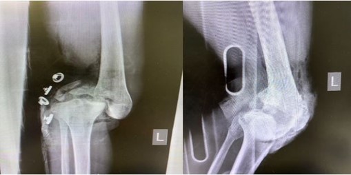
Figure 1. Pre-operative AP and lateral radiographs of the left knee showing the comminuted intra-articular distal femur fracture (open type C3).
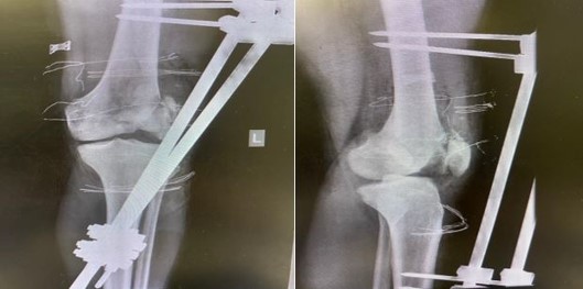
Figure 2. Post-operative images after first-stage surgery with a temporary unilateral external fixator spanning the femur and tibia.
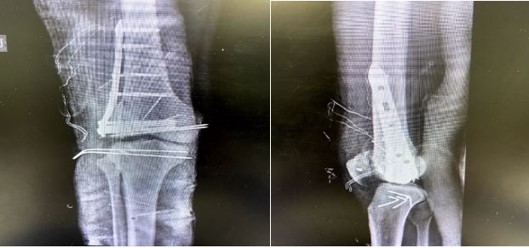
Figure 3. Plain radiographs at 8 weeks after definitive surgery, demonstrating the plate fixation in place and early callus formation at the fracture site.
Full-Text: (46 Views)
ABSTRACT
Open distal femur fractures are uncommon but challenging injuries, often resulting from high-energy trauma and associated with substantial risks of infection, non:union:, and functional impairment. Severe injuries with bone loss and concomitant disruption of the extensor mechanism present additional reconstructive challenges. We present the case of a 20-year-old man with an open, highly comminuted AO/OTA 33-C3 distal femur fracture with extensive soft-tissue injury, including complete patellar tendon rupture, lateral collateral ligament injury, and lateral retinacular disruption. The patient was managed using a staged approach consisting of initial irrigation and debridement with temporary external fixation, followed six weeks later by definitive open reduction and internal fixation using a distal femur locking plate, cancellous autologous iliac crest bone graft, and polypropylene mesh containment, together with repair of the extensor mechanism and lateral collateral ligament.
At 8 weeks after definitive fixation, the knee was clinically stable, radiographs demonstrated maintained alignment, stable fixation, and early callus formation, and range of motion had improved to approximately 0–120°. The patient was able to perform a straight-leg raise without extensor lag. No infection, hardware failure, or loss of ligamentous repair was observed during the reported follow-up.
Polypropylene mesh may be a useful adjunct for containment of autologous bone graft in selected distal femoral metaphyseal defects. In complex open distal femur fractures with extensor mechanism disruption, staged management, meticulous debridement, stable fixation, management of bone loss, and restoration of the extensor mechanism are important components of treatment.
Keywords: Distal femur fracture; Open fracture; Mesh augmentation; Bone loss; Patellar tendon rupture; Extensor mechanism
Open distal femur fractures are uncommon but challenging injuries, often resulting from high-energy trauma and associated with substantial risks of infection, non:union:, and functional impairment. Severe injuries with bone loss and concomitant disruption of the extensor mechanism present additional reconstructive challenges. We present the case of a 20-year-old man with an open, highly comminuted AO/OTA 33-C3 distal femur fracture with extensive soft-tissue injury, including complete patellar tendon rupture, lateral collateral ligament injury, and lateral retinacular disruption. The patient was managed using a staged approach consisting of initial irrigation and debridement with temporary external fixation, followed six weeks later by definitive open reduction and internal fixation using a distal femur locking plate, cancellous autologous iliac crest bone graft, and polypropylene mesh containment, together with repair of the extensor mechanism and lateral collateral ligament.
At 8 weeks after definitive fixation, the knee was clinically stable, radiographs demonstrated maintained alignment, stable fixation, and early callus formation, and range of motion had improved to approximately 0–120°. The patient was able to perform a straight-leg raise without extensor lag. No infection, hardware failure, or loss of ligamentous repair was observed during the reported follow-up.
Polypropylene mesh may be a useful adjunct for containment of autologous bone graft in selected distal femoral metaphyseal defects. In complex open distal femur fractures with extensor mechanism disruption, staged management, meticulous debridement, stable fixation, management of bone loss, and restoration of the extensor mechanism are important components of treatment.
Keywords: Distal femur fracture; Open fracture; Mesh augmentation; Bone loss; Patellar tendon rupture; Extensor mechanism
1. Introduction
Distal femur fractures account for less than 1% of all fractures, with approximately 5–10% presenting as open injuries and typically resulting from high-energy mechanisms in young patients (1,2). Open distal femur fractures are associated with substantial risks of infection, non:union:, and post-traumatic knee stiffness (1,3). Associated injuries around the knee may include ligamentous disruption and extensor mechanism injuries (4,5,6). Failure to recognize and appropriately address patellar or quadriceps tendon disruption may adversely affect functional recovery (4,6). Severe open fractures with bone loss are commonly managed using staged protocols, including induced membrane (Masquelet) reconstruction and related techniques (7,8–10). Modified techniques using mesh or cages to contain bone graft have also been described (11,12). To our knowledge, few reports have described polypropylene mesh as a containment device for bone graft in an open distal femur fracture combined with simultaneous ligamentous and extensor mechanism repair. We present this case and its early clinical and radiographic outcome.
2. Case Presentation
A 20-year-old man was brought to the emergency department after a motorcycle rollover accident. He sustained an open fracture of the left distal femur with a large wound over the anterolateral aspect of the knee. Examination revealed an approximately 10-cm laceration with exposed comminuted distal femoral bone fragments. The knee was grossly deformed and unstable, with virtually no active range of motion because of pain and mechanical disruption. Distal neurovascular examination was intact, with palpable pedal pulses and normal motor and sensory function in the foot. No other significant injuries were identified, and the patient had no relevant medical history. Radiographs of the left femur and knee demonstrated a highly comminuted intra-articular distal femur fracture with metaphyseal fragmentation and displacement (Figure 1). The fracture was classified as AO/OTA 33-C3, representing a complete articular fracture with a multifragmentary pattern.
The radiographs also suggested associated soft-tissue injury, including patella alta, consistent with patellar tendon disruption, and an avulsion fragment at the lateral femoral condyle suggestive of lateral collateral ligament injury. Following initial resuscitation and administration of broad-spectrum antibiotics according to the open-fracture protocol, the patient underwent emergency surgery. During the first stage, the wound was extensively irrigated with approximately 9 L of normal saline, and all contaminated and devitalized tissue was debrided. Knee arthrotomy was performed for joint lavage and removal of debris and fracture fragments. Intraoperatively, the patellar tendon was found to be completely ruptured from its tibial tubercle insertion; the lateral collateral ligament and lateral patellar retinaculum were also disrupted.
Because of the degree of contamination and comminution, definitive internal fixation was deferred and damage-control stabilization was performed. A uniplanar external fixator was applied across the knee to stabilize the femur and tibia in an aligned position (Figure 2). Definitive tendon and ligament repair was postponed; the extensor mechanism was provisionally approximated, and the wound was packed with moist sterile dressings. The limb was elevated and intravenous antibiotics were continued. The patient tolerated the procedure without evidence of acute compartment syndrome or vascular compromise. Six weeks later, after improvement of the soft-tissue condition and in the absence of clinical signs of infection, the patient underwent the second-stage procedure. The external fixator was removed, and formal open reduction and internal fixation of the distal femur was performed through a lateral approach. The fracture was reduced and stabilized with a stainless-steel distal femur locking compression plate contoured to the lateral condyle. Locking screws were placed proximally and distally to secure the major fracture fragments. A large metaphyseal bone defect remained because of bone loss. Cancellous autograft harvested from the ipsilateral iliac crest was packed into the defect. A standard surgical polypropylene mesh was then wrapped around the comminuted area to contain the graft and fracture fragments and was sutured to the adjacent intact periosteum and plate for reinforcement. During the same procedure, the associated soft-tissue injuries were addressed. The patellar tendon was reattached to the tibial tubercle using multiple strong nonabsorbable FiberWire sutures passed through transosseous tunnels and supplemented with two suture anchors. The lateral collateral ligament was repaired with a suture anchor at its femoral insertion, and the lateral retinaculum was repaired. The popliteal artery was visualized and found to be patent; therefore, vascular repair was not required. After thorough irrigation, closed-suction drains were placed, and the wound was closed in layers over the mesh and plate. Postoperatively, the knee was maintained in full extension using a hinged brace locked for 2 weeks to protect the patellar tendon repair. Distal pulses and neurological status remained normal. Gentle range-of-motion exercises, including passive flexion and active extension, were initiated under supervision at 2 weeks after surgery once satisfactory wound healing had been achieved.
At 8 weeks after definitive surgery, the knee was clinically stable without varus or valgus laxity. Range of motion had improved from approximately 0–30° in the early postoperative period to approximately 0–120°. The patient was able to perform a straight-leg raise without extensor lag. Follow-up radiographs demonstrated maintained alignment, stable hardware, and early callus formation around the fracture site (Figure 3). No infection, hardware loosening, or failure of the ligament repairs was observed during the reported follow-up period.
2. Case Presentation
A 20-year-old man was brought to the emergency department after a motorcycle rollover accident. He sustained an open fracture of the left distal femur with a large wound over the anterolateral aspect of the knee. Examination revealed an approximately 10-cm laceration with exposed comminuted distal femoral bone fragments. The knee was grossly deformed and unstable, with virtually no active range of motion because of pain and mechanical disruption. Distal neurovascular examination was intact, with palpable pedal pulses and normal motor and sensory function in the foot. No other significant injuries were identified, and the patient had no relevant medical history. Radiographs of the left femur and knee demonstrated a highly comminuted intra-articular distal femur fracture with metaphyseal fragmentation and displacement (Figure 1). The fracture was classified as AO/OTA 33-C3, representing a complete articular fracture with a multifragmentary pattern.
The radiographs also suggested associated soft-tissue injury, including patella alta, consistent with patellar tendon disruption, and an avulsion fragment at the lateral femoral condyle suggestive of lateral collateral ligament injury. Following initial resuscitation and administration of broad-spectrum antibiotics according to the open-fracture protocol, the patient underwent emergency surgery. During the first stage, the wound was extensively irrigated with approximately 9 L of normal saline, and all contaminated and devitalized tissue was debrided. Knee arthrotomy was performed for joint lavage and removal of debris and fracture fragments. Intraoperatively, the patellar tendon was found to be completely ruptured from its tibial tubercle insertion; the lateral collateral ligament and lateral patellar retinaculum were also disrupted.
Because of the degree of contamination and comminution, definitive internal fixation was deferred and damage-control stabilization was performed. A uniplanar external fixator was applied across the knee to stabilize the femur and tibia in an aligned position (Figure 2). Definitive tendon and ligament repair was postponed; the extensor mechanism was provisionally approximated, and the wound was packed with moist sterile dressings. The limb was elevated and intravenous antibiotics were continued. The patient tolerated the procedure without evidence of acute compartment syndrome or vascular compromise. Six weeks later, after improvement of the soft-tissue condition and in the absence of clinical signs of infection, the patient underwent the second-stage procedure. The external fixator was removed, and formal open reduction and internal fixation of the distal femur was performed through a lateral approach. The fracture was reduced and stabilized with a stainless-steel distal femur locking compression plate contoured to the lateral condyle. Locking screws were placed proximally and distally to secure the major fracture fragments. A large metaphyseal bone defect remained because of bone loss. Cancellous autograft harvested from the ipsilateral iliac crest was packed into the defect. A standard surgical polypropylene mesh was then wrapped around the comminuted area to contain the graft and fracture fragments and was sutured to the adjacent intact periosteum and plate for reinforcement. During the same procedure, the associated soft-tissue injuries were addressed. The patellar tendon was reattached to the tibial tubercle using multiple strong nonabsorbable FiberWire sutures passed through transosseous tunnels and supplemented with two suture anchors. The lateral collateral ligament was repaired with a suture anchor at its femoral insertion, and the lateral retinaculum was repaired. The popliteal artery was visualized and found to be patent; therefore, vascular repair was not required. After thorough irrigation, closed-suction drains were placed, and the wound was closed in layers over the mesh and plate. Postoperatively, the knee was maintained in full extension using a hinged brace locked for 2 weeks to protect the patellar tendon repair. Distal pulses and neurological status remained normal. Gentle range-of-motion exercises, including passive flexion and active extension, were initiated under supervision at 2 weeks after surgery once satisfactory wound healing had been achieved.
At 8 weeks after definitive surgery, the knee was clinically stable without varus or valgus laxity. Range of motion had improved from approximately 0–30° in the early postoperative period to approximately 0–120°. The patient was able to perform a straight-leg raise without extensor lag. Follow-up radiographs demonstrated maintained alignment, stable hardware, and early callus formation around the fracture site (Figure 3). No infection, hardware loosening, or failure of the ligament repairs was observed during the reported follow-up period.
Figure 1. Pre-operative AP and lateral radiographs of the left knee showing the comminuted intra-articular distal femur fracture (open type C3).
Figure 2. Post-operative images after first-stage surgery with a temporary unilateral external fixator spanning the femur and tibia.
Figure 3. Plain radiographs at 8 weeks after definitive surgery, demonstrating the plate fixation in place and early callus formation at the fracture site.
3. Discussion
Open distal femur fractures are complex injuries associated with substantial risks of infection and disability (1-3). Concomitant complete disruption of the extensor mechanism is uncommon but has been reported (4). Early recognition of associated tendon and ligament injuries is important because these injuries can substantially influence postoperative knee function (4,6). For severe open fractures, staged treatment can facilitate aggressive initial debridement, temporary stabilization, and subsequent definitive reconstruction after improvement of the soft-tissue envelope (7). Temporary spanning external fixation is commonly used as a damage-control strategy, while definitive fixation with a locking plate is an established option for comminuted distal femur fractures (1,13,14). Bone loss remains a major reconstructive challenge. The induced membrane (Masquelet) technique is widely used for segmental and metaphyseal bone defects (8–10). Kalantar et al. described the use of polypropylene mesh in a modified Masquelet technique for open distal femur fractures (11), while titanium cages have also been reported for large segmental defects (12).
In the present case, polypropylene mesh was used as a containment device for cancellous autograft within a metaphyseal defect. Its use may offer a simple and relatively inexpensive means of maintaining graft containment in selected defects; however, the present single case cannot establish comparative effectiveness or superiority over other reconstructive strategies. Restoration of the extensor mechanism is essential for functional recovery after severe knee trauma (4-6,15). Synthetic mesh and other augmentation techniques have been described for complex tendon repairs (16,17). In this case, transosseous sutures combined with suture anchors were used to obtain a robust patellar tendon repair, permitting supervised early rehabilitation. At the reported 8-week follow-up, the patient had a stable knee, approximately 0–120° of motion, and a straight-leg raise without extensor lag. Radiographs demonstrated maintained alignment, stable fixation, and early callus formation. No infection or implant-related complication was observed during this short follow-up.
These findings represent a favorable early result, but longer follow-up is necessary to determine fracture :union:, durability of the soft-tissue repairs, and functional outcome.
The principal limitation of this report is its single-case design and short follow-up period. In addition, the experience described here does not permit determination of whether polypropylene mesh provides advantages over established bone-defect reconstruction techniques. Further clinical studies are required to evaluate the safety, durability, and reproducibility of this approach.
4. Conclusion
Staged management of severe open distal femur fractures with associated ligamentous and tendon injuries may provide satisfactory early clinical and functional outcomes. Polypropylene mesh may be considered as an adjunct for containing autologous bone graft in selected metaphyseal bone defects. Meticulous debridement, temporary stabilization during the acute phase, definitive fixation after soft-tissue recovery, appropriate management of bone loss, and restoration of the extensor mechanism are important components of treatment in these complex injuries.
5. Declarations
5.1 Acknowledgments
The authors would like to express their sincere gratitude to the medical and nursing staff of Mousavi Hospital Complex, Zanjan University of Medical Sciences, for their valuable assistance in the management and care of the patient.
The authors also gratefully acknowledge the support and assistance of the Research and Technology Deputy of Zanjan University of Medical Sciences during the preparation of this manuscript.
5.2 Ethical Considerations
Written informed consent was obtained from the patient for publication of this case report and any accompanying images.
5.3 Authors' Contributions
S.Sh: conception and design of the study, clinical management, data collection, manuscript drafting, and critical revision of the manuscript. A.N: clinical management, data collection, interpretation of clinical and radiographic findings, and critical revision of the manuscript. S.R.A: data collection, literature review, manuscript drafting, and critical revision of the manuscript. All authors read and approved the final version of the manuscript.
5.4 Conflict of Interest
The authors declare that they have no conflict of interest.
5.5 Fund or Financial Support
No specific funding was received for this work.
5.6 Using Artificial Intelligence Tools (AI Tools)
AI tools were used for language editing and proofreading. All scientific content was reviewed and approved by the authors.
Open distal femur fractures are complex injuries associated with substantial risks of infection and disability (1-3). Concomitant complete disruption of the extensor mechanism is uncommon but has been reported (4). Early recognition of associated tendon and ligament injuries is important because these injuries can substantially influence postoperative knee function (4,6). For severe open fractures, staged treatment can facilitate aggressive initial debridement, temporary stabilization, and subsequent definitive reconstruction after improvement of the soft-tissue envelope (7). Temporary spanning external fixation is commonly used as a damage-control strategy, while definitive fixation with a locking plate is an established option for comminuted distal femur fractures (1,13,14). Bone loss remains a major reconstructive challenge. The induced membrane (Masquelet) technique is widely used for segmental and metaphyseal bone defects (8–10). Kalantar et al. described the use of polypropylene mesh in a modified Masquelet technique for open distal femur fractures (11), while titanium cages have also been reported for large segmental defects (12).
In the present case, polypropylene mesh was used as a containment device for cancellous autograft within a metaphyseal defect. Its use may offer a simple and relatively inexpensive means of maintaining graft containment in selected defects; however, the present single case cannot establish comparative effectiveness or superiority over other reconstructive strategies. Restoration of the extensor mechanism is essential for functional recovery after severe knee trauma (4-6,15). Synthetic mesh and other augmentation techniques have been described for complex tendon repairs (16,17). In this case, transosseous sutures combined with suture anchors were used to obtain a robust patellar tendon repair, permitting supervised early rehabilitation. At the reported 8-week follow-up, the patient had a stable knee, approximately 0–120° of motion, and a straight-leg raise without extensor lag. Radiographs demonstrated maintained alignment, stable fixation, and early callus formation. No infection or implant-related complication was observed during this short follow-up.
These findings represent a favorable early result, but longer follow-up is necessary to determine fracture :union:, durability of the soft-tissue repairs, and functional outcome.
The principal limitation of this report is its single-case design and short follow-up period. In addition, the experience described here does not permit determination of whether polypropylene mesh provides advantages over established bone-defect reconstruction techniques. Further clinical studies are required to evaluate the safety, durability, and reproducibility of this approach.
4. Conclusion
Staged management of severe open distal femur fractures with associated ligamentous and tendon injuries may provide satisfactory early clinical and functional outcomes. Polypropylene mesh may be considered as an adjunct for containing autologous bone graft in selected metaphyseal bone defects. Meticulous debridement, temporary stabilization during the acute phase, definitive fixation after soft-tissue recovery, appropriate management of bone loss, and restoration of the extensor mechanism are important components of treatment in these complex injuries.
5. Declarations
5.1 Acknowledgments
The authors would like to express their sincere gratitude to the medical and nursing staff of Mousavi Hospital Complex, Zanjan University of Medical Sciences, for their valuable assistance in the management and care of the patient.
The authors also gratefully acknowledge the support and assistance of the Research and Technology Deputy of Zanjan University of Medical Sciences during the preparation of this manuscript.
5.2 Ethical Considerations
Written informed consent was obtained from the patient for publication of this case report and any accompanying images.
5.3 Authors' Contributions
S.Sh: conception and design of the study, clinical management, data collection, manuscript drafting, and critical revision of the manuscript. A.N: clinical management, data collection, interpretation of clinical and radiographic findings, and critical revision of the manuscript. S.R.A: data collection, literature review, manuscript drafting, and critical revision of the manuscript. All authors read and approved the final version of the manuscript.
5.4 Conflict of Interest
The authors declare that they have no conflict of interest.
5.5 Fund or Financial Support
No specific funding was received for this work.
5.6 Using Artificial Intelligence Tools (AI Tools)
AI tools were used for language editing and proofreading. All scientific content was reviewed and approved by the authors.
Type of Study: Case Report Article |
Subject:
Clinical Medicine
Received: 2026/01/10 | Accepted: 2026/03/17 | Published: 2026/06/29
Received: 2026/01/10 | Accepted: 2026/03/17 | Published: 2026/06/29
References
1. Maley DK, Singh R, Khiyani R, Kaur K. Evaluation of results of open distal femur fractures with primary fixation and antibiotic impregnated collagen. Chin J Traumatol. 2019;22(6):328-32. [DOI:10.1016/j.cjtee.2019.08.005] [PMID] [PMCID]
2. Ehlinger M, Ducrot G, Adam P, Bonnomet F. Distal femur fractures. Surgical techniques and a review of the literature. Orthop Traumatol Surg Res. 2013;99(3):353-60. [DOI:10.1016/j.otsr.2012.10.014] [PMID]
3. Zhu C, Zhang J, Li J, Zhao K, Meng H, Zhu Y, et al. Incidence and predictors of surgical site infection after distal femur fractures treated by open reduction and internal fixation: a prospective single-center study. BMC Musculoskelet Disord. 2021;22:258. [DOI:10.1186/s12891-021-04132-9] [PMID] [PMCID]
4. Irigoyen MC, Bidolegui F, Pereira S. Fractures of the distal femur associated with a complete quadricipital tendon injury: report of two cases. Rev Asoc Argent Ortop Traumatol. 2022;87(2):238-45. [DOI:10.15417/issn.1852-7434.2022.87.2.1502]
5. Clayton RA, Court-Brown CM. The epidemiology of musculoskeletal tendinous and ligamentous injuries. Injury. 2008;39(12):1338-44. [DOI:10.1016/j.injury.2008.06.021] [PMID]
6. Siwek CW, Rao JP. Ruptures of the extensor mechanism of the knee joint. J Bone Joint Surg Am. 1981;63(6):932-937. [DOI:10.2106/00004623-198163060-00010]
7. Dugan TR, Hubert MG, Siska PA, Pape HC, Tarkin IS. Open supracondylar femur fractures with bone loss in the polytraumatized patient: timing is everything. Injury. 2013;44(12):1826-31. [DOI:10.1016/j.injury.2013.03.018] [PMID]
8. Masquelet AC, Fitoussi F, Begue T, Muller GP. Reconstruction of the long bones by the induced membrane and spongy autograft. Ann Chir Plast Esthet. 2000;45(3):346-53.
9. Taylor BC, Hancock J, Zitzke R, Castaneda J. Treatment of bone loss with the induced membrane technique: techniques and outcomes. J Orthop Trauma. 2015;29(12):554-57. [DOI:10.1097/BOT.0000000000000338] [PMID]
10. Karger C, Kishi T, Schneider L, Fitoussi F, Masquelet AC. Treatment of posttraumatic bone defects by the induced membrane technique. Orthop Traumatol Surg Res. 2012;98(1):97-102. [DOI:10.1016/j.otsr.2011.11.001] [PMID]
11. Kalantar SH, Saffar H, Hoveidaei AH. Bone reconstruction with modified Masquelet technique in open distal femoral fractures: a case series. BMC Musculoskelet Disord. 2024;25(1):26. [DOI:10.1186/s12891-023-07091-5] [PMID] [PMCID]
12. Ma XY, Yuan H, Cui D, Liu B, Han TY, Yu HL, et al. Management of segmental defects post open distal femur fracture using a titanium cage combined with the Masquelet technique: a single-centre report of 23 cases. Injury. 2023;54(12):111130. [DOI:10.1016/j.injury.2023.111130] [PMID]
13. Vosoughi F, Vosoughi F, Kalantar SH. Open knee dislocation, triple intra-articular fractures and patellar tendon rupture: a case report of a knee disaster treated with aggressive irrigation/debridement, early anatomic reduction and internal fixation. BMC Musculoskelet Disord. 2022;23:431. [DOI:10.1186/s12891-022-05268-y] [PMID] [PMCID]
14. Henderson CE, Lujan TJ, Kuhl LL, Bottlang M, Fitzpatrick DC, Marsh JL. Midshaft and distal femur fracture fixation: a biomechanical comparison of locked plating and intramedullary nailing. J Orthop Trauma. 2010;24(10):567-575.
15. Jaramillo Quiceno GA, Sarmiento Riveros PA, Arias Perez RD, Soto Gomez MP, Ramirez AO. Augmentation in the repair of traumatic patellar tendon ruptures: a novel mechanical and biological construct (Technical note). J ISAKOS. 2023;8(2):122-127. [DOI:10.1016/j.jisako.2022.10.003] [PMID]
16. Nodzo SR, Rachala SR. Polypropylene mesh augmentation for complete quadriceps rupture after total knee arthroplasty. Knee. 2016;23(1):177-180. [DOI:10.1016/j.knee.2015.09.007] [PMID]
17. Powell C, Sanders K, Huang N, Colón LF, Norton C. Distal femoral replacement and extensor mechanism repair reinforced with synthetic mesh for distal femur fracture with patellar ligament avulsion: a case report. Arthroplasty Today. 2022;16:31-38. [DOI:10.1016/j.artd.2022.04.001] [PMID] [PMCID]
Send email to the article author
| Rights and permissions | |
 |
This work is licensed under a Creative Commons Attribution-NonCommercial 4.0 International License. |
Copyright Policy
Journal of Advances in Medical and Biomedical Research by Zanjan University of Medical Sciences is licensed under CC BY-NC 4.0