
Sat, Nov 14, 2026
[Archive]
Volume 34, Issue 4 (July & August 2026)
J Adv Med Biomed Res 2026, 34(4): 380-387 |
Back to browse issues page



Download citation:
BibTeX | RIS | EndNote | Medlars | ProCite | Reference Manager | RefWorks
Send citation to:


BibTeX | RIS | EndNote | Medlars | ProCite | Reference Manager | RefWorks
Send citation to:
Al-Tememi M M, Al-Badr R J, Al-Khafaji A M. Antibacterial and Biofilm Reduction Effects of Totarol versus Calcium Hydroxide against Clinical Isolated Enterococcus faecalis: An In Vitro and Ex Vivo Assessment. J Adv Med Biomed Res 2026; 34 (4) :380-387
URL: http://journal.zums.ac.ir/article-1-7933-en.html
URL: http://journal.zums.ac.ir/article-1-7933-en.html


1- Ahl al-Bayt University, College of dentistry, Karbala, Iraq , mustafa.mkj94@gmail.com
2- Ahl al-Bayt University, College of dentistry, Karbala, Iraq
2- Ahl al-Bayt University, College of dentistry, Karbala, Iraq
Keywords: Enterococcus faecalis, Biofilms, Root Canal Therapy, Calcium Hydroxide, Antibacterial Agents, Endodontics
Full-Text [PDF 336 kb]
(98 Downloads)
| Abstract (HTML) (869 Views)
1. Introduction
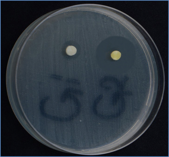
Figure 1. E. faecalis sensitivity to Totarol and Ca (OH)2 where the mean sensitivity was 38.26 and 15.69 respectively. (Prepared by Authors, 2026).
Table 2. Agar diffusion zone (Independent t-test).
Table 3. CFU before and after treatment (Paired t-test).
Note: SE calculated as SD/√n
Table 4. Statistical t-test summary (Paired + Independent).
Table 5. One-way ANOVA for post-treatment CFU.
Table 6. Tukey multiple-comparison output.
Full-Text: (52 Views)
1. Introduction
Persistent endodontic infections pose a significant challenge in clinical practice (1). Persistent endodontic infections are polymicrobial in nature; however, E. faecalis has been detected in 24–77% of failed root canal treatments, making it one of the most clinically relevant pathogens associated with retreatment cases (2). Its survival is attributed to resistance to nutrient deprivation, tolerance to alkaline stress, and the ability to penetrate dentinal tubules and form structured biofilms. The increasing emergence of vancomycin-resistant E. faecalis (VRE) strains further highlights the need for alternative antimicrobial strategies in endodontics (3). Totarol has demonstrated reported MIC values ranging from 0.25–4 µg/mL against Gram-positive bacteria in previous microbiological studies, suggesting potent membrane-targeting and electron-transport inhibitory activity (3). Despite these promising antimicrobial characteristics, its application as an intracanal medicament against mature endodontic biofilms remains insufficiently investigated. These traits enable E. faecalis to persist in root canals despite conventional chemomechanical preparation and irrigation (4).
Calcium hydroxide has long been the standard intracanal medicament because of its high pH and bactericidal activity mediated by hydroxyl ions (5). However, its effectiveness against E. faecalis is often limited due to dentin buffering and the bacterium's intrinsic alkaline tolerance (6-7).
Totarol is a naturally occurring phenolic diterpene derived from Podocarpus species (3). It has demonstrated potent activity against several Gram‑positive bacteria through mechanisms involving disruption of bacterial membranes, interference with metabolic pathways, and inhibition of electron transport (3). Unlike Ca (OH)2, Totarol is lipophilic, which may allow enhanced penetration into biofilm matrices and dentinal tubules. This property suggests a potential advantage in eradicating bacteria protected within biofilms. The present study was designed to compare the antibacterial and antibiofilm effects of Totarol (Bioactive Totarol Ltd., New Zealand) and Ca (OH)2 (Metapaste, Meta Biomed. Korea) using clinical E. faecalis isolates in both in vitro assays and an ex vivo 21‑day root canal biofilm model. The null hypothesis was that no significant difference exists between Totarol and Ca (OH)2 in their antimicrobial effectiveness against E. faecalis.
2. Materials and Methods
2.1 Bacterial Identification
Patients requiring endodontic treatment for pulpal necrosis associated with periapical lesions were recruited from the Karbala Dentistry Center, Iraq. The diagnosis was established based on clinical examination and radiographic findings obtained in the Diagnostic Unit.
Inclusion criteria were: (i) patients aged 25–45 years; (ii) single-rooted teeth diagnosed with pulpal necrosis and periapical pathology; and (iii) the presence of clinical symptoms indicative of endodontic infection. Exclusion criteria included: (i) current or recent use of systemic antibiotics; (ii) teeth with extensive coronal destruction that precluded proper rubber dam isolation; and (iii) teeth presenting with root or crown cracks. Under aseptic conditions, the tooth was isolated with a rubber dam, and the tooth and surrounding area were disinfected with 10% povidone–iodine. Access cavity preparation was done in two steps: first, a sterile carbide fissure bur was used to remove all caries and coronal restorations, followed by disinfection; second, a low-speed round carbide bur was used to remove the roof of the pulp chamber and complete cleaning. A new sterile #20 file was then placed in the root canal to the working length (confirmed radiographically), rotated twice clockwise, and immediately transferred into a tube containing sterile Amies transport medium (8). Because Enterococcus faecalis is a facultative anaerobe, samples were transported under both aerobic and anaerobic conditions to maximize bacterial recovery and then isolated within two hours.
Experiments were performed using a single confirmed clinical isolate of Enterococcus faecalis. All assays were repeated to ensure experimental reproducibility.
2.2 Determination of minimum inhibitory concentration (MIC) and minimum bactericidal concentration (MBC) for Totarol
MIC values for Totarol were determined using a broth dilution method with decreasing concentrations according to the Clinical and Laboratory Standards Institute guidelines (9). Tubes containing Mueller–Hinton broth were inoculated with standardized bacterial suspensions and incubated at 37°C for 24 hours under aerobic conditions. MIC was defined as the lowest concentration of Totarol that prevented visible turbidity. For MBC determination, aliquots from tubes without visible growth were sub‑cultured into agar plates (10). The lowest concentration yielding no bacterial growth on agar was considered the MBC. MIC/MBC determination for Ca (OH)₂ was not performed because its paste formulation and alkaline antimicrobial mechanism are not compatible with standardized broth dilution methods.
2.3 Agar Diffusion Assay
Muller‑Hinton agar plates were inoculated with standardized 0.5 McFarland suspensions of E. faecalis to create a uniform bacterial lawn (11). Sterile wells were prepared in the agar each with a diameter of 6 mm and a depth of 4 mm, were created in each agar plate using agar punch and filled with Totarol at the MBC concentration determined in broth dilution testing and Ca (OH)2 paste. The plates were incubated aerobically at 37 °C for 24 hours, and the diameter of the inhibition zones around each well was measured in millimeters using an electronic Vernier caliper (12).
2.4 Agar Diffusion Assay
Extracted human single-rooted teeth with fully formed apices were selected. The teeth selected for this study were first examined carefully, and any teeth showing cracks or defects in the cementum were excluded. Extracted teeth were stored in sterile saline at room temperature until use to prevent dehydration. The crowns were removed to obtain root segments of about 12 mm in length. A size 15 K-file was used to confirm apical patency and create a glide path. The working length was set 1 mm short of the point where the file tip became visible at the apical foramen. Specimens were randomly allocated into experimental groups (n = 10 per group). Root canals were shaped with a crown-down technique using the ProTaper Next files (X1–X4). Final irrigation for 1 minute was done with 17% EDTA and 2.5% NaOCl to remove debris and the smear layer, then the canals were rinsed with sterile distilled water. After instrumentation, the apical foramina of all roots were sealed with a flowable composite resin. The specimens were then placed vertically in silicone impression blocks. Before inoculation, they were sterilized in an autoclave at 121°C and 15 psi for 30 minutes. Resin integrity and apical sealing were verified after sterilization to ensure structural stability.
2.5 Biofilm Development (21‑Day Model)
One milliliter of bacterial isolates grown in Brain–heart infusion broth (BHI) for 24 hours was transferred into 5 mL of fresh Brain Heart Infusion broth to prepare the bacterial suspension. Using sterile 1-mL insulin syringes, the suspension was carefully injected into each root canal, taking care to avoid any overflow. A size 15 sterile K-file was then introduced with a single in-and-out movement to help spread the bacteria along the full length of the canal. After inoculation, the roots were stored in an incubator at 37°C for 21 days under aseptic conditions. All procedures were performed inside a laminar flow cabinet to minimize contamination risk. To maintain bacterial viability, fresh suspension was added every three days. Sterile broth-only controls were incubated in parallel to confirm absence of contamination during the biofilm development period.
2.6 Intracanal Medication
After biofilm maturation, specimens were randomly assigned into two groups: Totarol and Ca (OH)2. Totarol was applied inside the canals at its MBC concentration using 30-gauge syringe. Ca (OH)2 paste was placed using a Lentulo spiral according to manufacturer instructions in the canals of the comparator group After that, access cavities were sealed with temporary restorative material. All specimens were incubated at 37°C under aerobic conditions for an additional 3 days to allow interaction between medicaments and the biofilm. Totarol was dissolved in dimethyl sulfoxide (DMSO) to ensure complete solubility for intracanal application. A DMSO vehicle-control was tested separately and produced no measurable antibacterial effect under the experimental conditions. Each medicament was applied in the form consistent with its standard laboratory or commercial preparation to reflect realistic clinical handling conditions. The study aimed to compare functional antimicrobial performance rather than identical formulation viscosity.
A 3-day medication period was intentionally selected to evaluate short-term antimicrobial performance under standardized laboratory conditions.
2.7 Dentin Shavings Collection and CFU Enumeration
At the end of the medication period, dentin shavings were collected from the canal walls using sterile H file instruments (H-file, Dentsply Maillefer, USA) with 10 standardized vertical strokes. The shavings were transferred into tubes containing 1 mL of sterile transport medium and vortexed for 30 seconds at high speed to detach adherent bacteria from dentin. Serial dilutions were prepared and plated on agar. After incubation, CFU were counted and expressed as CFU ×103. Baseline and post‑medication CFU counts were recorded for each group.
2.8 Statistical Analysis
All experimental data were analyzed using SPSS software (v. 26). Normality was verified prior to evaluation. Paired t-tests were used to evaluate within-group changes (before vs after medication). Independent t-tests were applied for direct comparison between Totarol and Ca (OH)₂ groups. One-way ANOVA followed by Tukey HSD post-hoc testing was used to confirm overall differences among experimental conditions. Data were expressed as mean ± SD, and statistical significance was set at p < 0.05 (highly significant when p < 0.0001).
3. Result
Statistical analysis demonstrated a pronounced reduction in bacterial load following totarol treatment relative to baseline. Raw CFU values were consistent across specimens and fell within a narrow experimental range, supporting measurement reliability. Confidence interval analysis confirmed the statistical robustness of the observed differences between groups. MIC and MBC testing were performed in triplicate to ensure reproducibility. Agar diffusion testing followed standardized well-diameter methodology; identical well dimensions were maintained across all groups to ensure valid comparative interpretation. The proximity between MIC and MBC values suggests strong bactericidal activity, although time-kill dynamics were not evaluated in this study. Data distribution normality was verified prior to parametric testing, and confidence interval analysis supported the statistical reliability of the observed reductions.
3.1 Identification of isolated bacteria
Colonies grew well on blood agar and showed no hemolysis. They were sized 1.0–1.5 mm and had a circular, smooth, raised edge. E. faecalis grows on Pfizer agar, resulting in a black colony radius. The VITEK 2 system confirmed identification with ≥99% confidence (Table 1).
Table 1. VITEK 2 identification results for Enterococcus faecalis isolates.
| Identification Information | Analysis Time: 3.10 hours | Status: Final | |||||||||||
| Selected Organism | 99% Probability Enterococcus faecalis Bionumber: 116002765773431 |
||||||||||||
| ID Analysis messages | |||||||||||||
| 2 | AMY | + | 4 | PIPLC | - | 5 | dXYL | - | 8 | ADH1 | + | ||
| 9 | BGAL | - | 11 | AGLU | - | 13 | APPA | - | 14 | CDEX | + | ||
| 15 | AspA | + | 16 | BGAR | - | 17 | AMAN | - | 19 | PHOS | - | ||
| 20 | LeuA | - | 23 | ProA | - | 24 | BGURr | - | 25 | AGAL | - | ||
| 26 | PyrA | + | 27 | BGUR | - | 28 | AlaA | + | 29 | TyrA | - | ||
| 30 | dSOR | + | 31 | URE | - | 32 | POLYB | + | 37 | dGAL | + | ||
| 38 | dRIB | + | 39 | ILATk | + | 42 | LAC | + | 44 | NAG | + | ||
| 45 | dMAL | + | 46 | BACI | + | 47 | NOVO | + | 50 | NC6.5 | + | ||
| 52 | dMAN | + | 53 | dMNE | + | 54 | MBdG | - | 56 | PUL | - | ||
| 57 | dRAF | - | 58 | O129R | - | 59 | SAL | + | 60 | SAC | + | ||
| 62 | dTRE | + | 63 | ADH2s | - | 64 | OPTO | + | |||||
3.2 MIC and MBC of Totarol
The MIC of Totarol against the clinical E. faecalis isolate was 11.25 µl/mL, and the MBC was 22.5 µl/mL. The units represent volumetric dilution of the totarol stock solution rather than mass concentration. MIC/MBC values for Ca (OH)₂ were not determined because its paste formulation and alkaline antimicrobial mechanism are not compatible with standardized broth dilution testing. The proximity between MIC and MBC values suggests strong bactericidal activity; however, bacterial viability dynamics and time-kill kinetics were not evaluated in the present study.
3.3 Agar Diffusion Test
Totarol produced a diffusion halo larger than Ca (OH)2 (Figure 1). The larger inhibition zone observed in the agar model reflects enhanced diffusion behavior under standardized laboratory conditions.
The independent t-test confirmed significance (p < 0.0001), indicating this difference is real and biologically meaningful as shown in Table 2.
3.4 Ex vivo 21-Day biofilm model
Totarol reduced bacterial load by approximately 97.8%, leaving minimal viable cells. Although a marked bacterial reduction was observed, complete eradication was not achieved within the experimental period. Ca (OH)2 achieved ~81.1%, significantly weaker in eliminating established biofilm. Paired t-tests yielded p < 0.0001, confirming reduction was statistically meaningful as shown in Table 3.
3.5 Significance testing (Comparative Statistics)
Statistical analysis confirmed significant differences between experimental groups, as detailed in Table 4 and Table 5.
The MIC of Totarol against the clinical E. faecalis isolate was 11.25 µl/mL, and the MBC was 22.5 µl/mL. The units represent volumetric dilution of the totarol stock solution rather than mass concentration. MIC/MBC values for Ca (OH)₂ were not determined because its paste formulation and alkaline antimicrobial mechanism are not compatible with standardized broth dilution testing. The proximity between MIC and MBC values suggests strong bactericidal activity; however, bacterial viability dynamics and time-kill kinetics were not evaluated in the present study.
3.3 Agar Diffusion Test
Totarol produced a diffusion halo larger than Ca (OH)2 (Figure 1). The larger inhibition zone observed in the agar model reflects enhanced diffusion behavior under standardized laboratory conditions.
The independent t-test confirmed significance (p < 0.0001), indicating this difference is real and biologically meaningful as shown in Table 2.
3.4 Ex vivo 21-Day biofilm model
Totarol reduced bacterial load by approximately 97.8%, leaving minimal viable cells. Although a marked bacterial reduction was observed, complete eradication was not achieved within the experimental period. Ca (OH)2 achieved ~81.1%, significantly weaker in eliminating established biofilm. Paired t-tests yielded p < 0.0001, confirming reduction was statistically meaningful as shown in Table 3.
3.5 Significance testing (Comparative Statistics)
Statistical analysis confirmed significant differences between experimental groups, as detailed in Table 4 and Table 5.
Figure 1. E. faecalis sensitivity to Totarol and Ca (OH)2 where the mean sensitivity was 38.26 and 15.69 respectively. (Prepared by Authors, 2026).
Table 2. Agar diffusion zone (Independent t-test).
| Group | N | Mean (mm) | SD | SE | ||
|
10 | 38.26 | 1.84 | 0.58 | ||
| Ca (OH)2 | 10 | 15.69 | 1.73 | 0.55 | ||
| Negative control | 10 | 0 | - | - |
Table 3. CFU before and after treatment (Paired t-test).
| Group | Stage | N | Mean CFU * 103 | SD | SE | ||||||
|
Before | 10 |
|
1.93 |
|
||||||
|
After | 10 | 1.4 |
|
|
||||||
| Ca (OH)2 | Before | 10 |
|
1.47 |
|
||||||
| Ca (OH)2 | After | 10 |
|
|
0.60 | ||||||
| Negative control (without medicament) | Before | 10 | 64.7 | 1.889 |
|
||||||
| Negative control (without medicament) | After | 10 | 59 | 1.945 | 0.615 |
Table 4. Statistical t-test summary (Paired + Independent).
| Comparison | Test Used | P-value | |||
|
|
|
|||
|
Paired t-test | <0.0001 | |||
|
|
<0.0001 |
|||
|
Paired t-test |
> 0.05 non-significant |
All comparisons revealed extremely significant differences as shown in Table 3. Although both medicaments lowered CFU, Totarol demonstrated the highest magnitude of reduction, proving more effective overall. Totarol yielded much lower final CFU counts, confirming superior bactericidal performance.
3.7 Tukey HSD post-Hoc comparison
post-hoc testing verifies that Totarol’s antibacterial effect is statistically superior as shown in Table 6. Totarol is the treatment responsible for the significance detected in ANOVA.
3.7 Tukey HSD post-Hoc comparison
post-hoc testing verifies that Totarol’s antibacterial effect is statistically superior as shown in Table 6. Totarol is the treatment responsible for the significance detected in ANOVA.
Table 5. One-way ANOVA for post-treatment CFU.
| Source | df | F-value | P-value | |||||
|
2 |
|
<0.0001 | |||||
|
27 |
- |
- |
|||||
Total |
29 |
- |
- |
|||||
Table 6. Tukey multiple-comparison output.
| Comparison | Mean Difference | Comparison |
| Totarol vs Ca (OH)2 | 10.3 | <0.0001 |
| Totarol vs Negative Control | 57.6 | < 0.0001 |
| CaOH vs Negative Control | 47.3 | < 0.001 |
4. Discussion
Within the limitations of the present laboratory model, Totarol demonstrated a greater antibacterial and biofilm-reducing effect compared with Ca (OH)₂ when tested against clinical E. faecalis isolates. Totarol consistently outperformed Ca (OH)2 in MIC/MBC testing, agar diffusion, and the ex vivo 21‑day biofilm model. The MIC and MBC values of Totarol further confirmed its potency. A MIC of 11.25 µl/ml and an MBC of 22.5 µl/ml indicate that Totarol can inhibit and kill E. faecalis at relatively low Concentrations (3) . The small difference between MIC and MBC suggests strong bactericidal activity; however, bacterial viability dynamics and time-kill kinetics were not evaluated in the present study. The agar diffusion results showed that Totarol produced a significantly larger inhibition zone (38.26 mm) compared to Ca (OH)2 (15.69 mm). This indicates enhanced diffusion capability, membrane permeability and surface efficacy (13). A broad inhibitory halo is suggestive of a compound that not only kills bacteria on contact, but also spreads effectively through dentinal pathways and surrounding medium.
Ca (OH)2, in contrast, demonstrated limited radial diffusion. Its antimicrobial action relies mainly on high alkalinity (pH ~12.5) (14), which is effective only when direct contact is achieved. This limitation explains its smaller inhibition zone and weaker ability to spread into deeper biofilm layers (15, 16). For clinical context, established intracanal disinfectants such as chlorhexidine and sodium hypochlorite have been reported to produce inhibition zones typically ranging between 30–40 mm in agar diffusion models. The inhibition zone observed for Totarol in the present study falls within this effective antimicrobial spectrum, suggesting comparable in vitro diffusion behavior. However, direct comparative testing with gold-standard irrigants was beyond the scope of this investigation (17, 18). The ex vivo biofilm model provided a clinically relevant scenario in which E. faecalis adopted a biofilm lifestyle inside dentinal tubules, consistent with established dentin infection models described in the endodontic literature (19). This model simulates intraradicular biofilm conditions associated with persistent endodontic infections. Totarol demonstrated a substantially greater bacterial reduction compared with Ca (OH)₂ in the biofilm model. Although a marked bacterial reduction was achieved, complete eradication was not observed within the experimental period.
E. faecalis is highly resistant to alkaline environments, survives in nutrient-deprived conditions, penetrates dentinal tubules, and forms strongly adherent biofilms (20). Ca (OH)2 kills bacteria mainly by hydroxyl ion release, and many E. faecalis strains develop defense mechanisms, including proton pumps and stress protein expression, allowing survival despite high pH (7). Several alternative intracanal agents, including chlorhexidine, triple antibiotic paste, and selected natural compounds such as propolis and essential oils, have demonstrated antibacterial activity against persistent endodontic pathogens. The present findings position totarol within this expanding group of candidate medicaments and highlight the importance of continued comparative research. The increasing prevalence of vancomycin-resistant E. faecalis, reported in approximately 15–30% of clinical isolates, further highlights the importance of exploring alternative antimicrobial strategies. Totarol has demonstrated activity against resistant Gram-positive strains in previous studies (3, 21, 22). Totarol’s antimicrobial activity has been associated with membrane disruption, oxidative stress induction, and interference with bacterial electron transport pathways (22). Previous microbiological investigations suggest that Totarol may also influence biofilm-associated gene expression and stress-response signaling. Although mechanistic assays were not performed in the present study, the pronounced reduction in viable bacterial counts is consistent with these proposed antimicrobial mechanisms (3, 22).
Although most mechanistic insights into Totarol originate from non-endodontic microbiological research, these studies provide foundational evidence of its antimicrobial behavior. Given the limited availability of endodontic-specific investigations, broader microbiological literature remains essential for interpreting its potential activity against oral biofilms. The ability of a medicament to penetrate deep dentinal layers and disrupt biofilm is critical for successful endodontic therapy (23). Although totarol is lipophilic, dentinal penetration depth was not directly measured in the present study. Imaging-based confirmation would be required to verify deeper penetration into dentinal tubules. The near-complete CFU elimination achieved by Totarol indicates that it may serve as a strong alternative or adjunct to Ca (OH)2, especially in retreatment and resistant infections. Potential clinical advantages of Totarol include: strong antibiofilm activity, stronger bactericidal effect, effective against highly resistant E. faecalis, and better diffusion than Ca (OH)2. This suggests that Totarol could enhance intracanal disinfection protocols and reduce treatment failure in persistent endodontic infections. However, clinical translation requires caution and further biological validation before routine clinical application. This study has several limitations. First, it was conducted in vitro and ex vivo, which does not fully replicate the complex conditions of the clinical environment, including host immune responses and mechanical factors. Second, the biofilm model used a mono-species E. faecalis biofilm, whereas clinical infections are often polymicrobial. Third, cytotoxicity, tissue compatibility, and pH changes during medication were not evaluated. Assessment of biological safety is essential before clinical application and should be addressed in future investigations. Finally, Totarol was evaluated in a laboratory solution format, and its performance in commercial formulations remains to be explored.
5. Conclusion
Within the limitations of this study, Totarol produced a substantially greater reduction in E. faecalis biofilm compared with Ca (OH)₂ (approximately 97.8% vs 81.1%; p < 0.0001). These findings demonstrate strong in vitro antibacterial performance against a key pathogen associated with persistent endodontic infections. However, laboratory results should not be interpreted as direct predictors of clinical success. Further investigation into biological safety, polymicrobial biofilm behavior, and clinical translation is required before routine application. These findings support continued exploration of novel antimicrobial strategies that may redefine future management of persistent endodontic infections.
6. Declarations
6.1 Acknowledgments
The authors would like to thank the staff of the laboratories involved in this study for their assistance and support during the experimental work.
6.2 Ethical Considerations
This study was approved by the scientific research committee at the College of Dentistry, University of Ahl Al-Bayt (Ref. 101 in 2026).
6.3 Authors' Contributions
Dr. Mustafa Mohammed Al-Tememi performed the experimental work and drafted the manuscript. Dr. Rafid Jihad Al-Badr supervised the study and critically reviewed the manuscript. Dr. Ahmed Mahdi Al-Khafaji contributed to the statistical analysis of the study. All authors read and approved the final version of the manuscript.
6.4 Conflict of Interest
The authors declare that there is no conflict of interest regarding the publication of this article.
6.5 Fund or Financial Support
This research received no specific grant from any funding agency in the public, commercial, or not-for-profit sectors.
6.6 Using Artificial Intelligence Tools (AI Tools)
The authors were not utilized AI Tools.
Within the limitations of the present laboratory model, Totarol demonstrated a greater antibacterial and biofilm-reducing effect compared with Ca (OH)₂ when tested against clinical E. faecalis isolates. Totarol consistently outperformed Ca (OH)2 in MIC/MBC testing, agar diffusion, and the ex vivo 21‑day biofilm model. The MIC and MBC values of Totarol further confirmed its potency. A MIC of 11.25 µl/ml and an MBC of 22.5 µl/ml indicate that Totarol can inhibit and kill E. faecalis at relatively low Concentrations (3) . The small difference between MIC and MBC suggests strong bactericidal activity; however, bacterial viability dynamics and time-kill kinetics were not evaluated in the present study. The agar diffusion results showed that Totarol produced a significantly larger inhibition zone (38.26 mm) compared to Ca (OH)2 (15.69 mm). This indicates enhanced diffusion capability, membrane permeability and surface efficacy (13). A broad inhibitory halo is suggestive of a compound that not only kills bacteria on contact, but also spreads effectively through dentinal pathways and surrounding medium.
Ca (OH)2, in contrast, demonstrated limited radial diffusion. Its antimicrobial action relies mainly on high alkalinity (pH ~12.5) (14), which is effective only when direct contact is achieved. This limitation explains its smaller inhibition zone and weaker ability to spread into deeper biofilm layers (15, 16). For clinical context, established intracanal disinfectants such as chlorhexidine and sodium hypochlorite have been reported to produce inhibition zones typically ranging between 30–40 mm in agar diffusion models. The inhibition zone observed for Totarol in the present study falls within this effective antimicrobial spectrum, suggesting comparable in vitro diffusion behavior. However, direct comparative testing with gold-standard irrigants was beyond the scope of this investigation (17, 18). The ex vivo biofilm model provided a clinically relevant scenario in which E. faecalis adopted a biofilm lifestyle inside dentinal tubules, consistent with established dentin infection models described in the endodontic literature (19). This model simulates intraradicular biofilm conditions associated with persistent endodontic infections. Totarol demonstrated a substantially greater bacterial reduction compared with Ca (OH)₂ in the biofilm model. Although a marked bacterial reduction was achieved, complete eradication was not observed within the experimental period.
E. faecalis is highly resistant to alkaline environments, survives in nutrient-deprived conditions, penetrates dentinal tubules, and forms strongly adherent biofilms (20). Ca (OH)2 kills bacteria mainly by hydroxyl ion release, and many E. faecalis strains develop defense mechanisms, including proton pumps and stress protein expression, allowing survival despite high pH (7). Several alternative intracanal agents, including chlorhexidine, triple antibiotic paste, and selected natural compounds such as propolis and essential oils, have demonstrated antibacterial activity against persistent endodontic pathogens. The present findings position totarol within this expanding group of candidate medicaments and highlight the importance of continued comparative research. The increasing prevalence of vancomycin-resistant E. faecalis, reported in approximately 15–30% of clinical isolates, further highlights the importance of exploring alternative antimicrobial strategies. Totarol has demonstrated activity against resistant Gram-positive strains in previous studies (3, 21, 22). Totarol’s antimicrobial activity has been associated with membrane disruption, oxidative stress induction, and interference with bacterial electron transport pathways (22). Previous microbiological investigations suggest that Totarol may also influence biofilm-associated gene expression and stress-response signaling. Although mechanistic assays were not performed in the present study, the pronounced reduction in viable bacterial counts is consistent with these proposed antimicrobial mechanisms (3, 22).
Although most mechanistic insights into Totarol originate from non-endodontic microbiological research, these studies provide foundational evidence of its antimicrobial behavior. Given the limited availability of endodontic-specific investigations, broader microbiological literature remains essential for interpreting its potential activity against oral biofilms. The ability of a medicament to penetrate deep dentinal layers and disrupt biofilm is critical for successful endodontic therapy (23). Although totarol is lipophilic, dentinal penetration depth was not directly measured in the present study. Imaging-based confirmation would be required to verify deeper penetration into dentinal tubules. The near-complete CFU elimination achieved by Totarol indicates that it may serve as a strong alternative or adjunct to Ca (OH)2, especially in retreatment and resistant infections. Potential clinical advantages of Totarol include: strong antibiofilm activity, stronger bactericidal effect, effective against highly resistant E. faecalis, and better diffusion than Ca (OH)2. This suggests that Totarol could enhance intracanal disinfection protocols and reduce treatment failure in persistent endodontic infections. However, clinical translation requires caution and further biological validation before routine clinical application. This study has several limitations. First, it was conducted in vitro and ex vivo, which does not fully replicate the complex conditions of the clinical environment, including host immune responses and mechanical factors. Second, the biofilm model used a mono-species E. faecalis biofilm, whereas clinical infections are often polymicrobial. Third, cytotoxicity, tissue compatibility, and pH changes during medication were not evaluated. Assessment of biological safety is essential before clinical application and should be addressed in future investigations. Finally, Totarol was evaluated in a laboratory solution format, and its performance in commercial formulations remains to be explored.
5. Conclusion
Within the limitations of this study, Totarol produced a substantially greater reduction in E. faecalis biofilm compared with Ca (OH)₂ (approximately 97.8% vs 81.1%; p < 0.0001). These findings demonstrate strong in vitro antibacterial performance against a key pathogen associated with persistent endodontic infections. However, laboratory results should not be interpreted as direct predictors of clinical success. Further investigation into biological safety, polymicrobial biofilm behavior, and clinical translation is required before routine application. These findings support continued exploration of novel antimicrobial strategies that may redefine future management of persistent endodontic infections.
6. Declarations
6.1 Acknowledgments
The authors would like to thank the staff of the laboratories involved in this study for their assistance and support during the experimental work.
6.2 Ethical Considerations
This study was approved by the scientific research committee at the College of Dentistry, University of Ahl Al-Bayt (Ref. 101 in 2026).
6.3 Authors' Contributions
Dr. Mustafa Mohammed Al-Tememi performed the experimental work and drafted the manuscript. Dr. Rafid Jihad Al-Badr supervised the study and critically reviewed the manuscript. Dr. Ahmed Mahdi Al-Khafaji contributed to the statistical analysis of the study. All authors read and approved the final version of the manuscript.
6.4 Conflict of Interest
The authors declare that there is no conflict of interest regarding the publication of this article.
6.5 Fund or Financial Support
This research received no specific grant from any funding agency in the public, commercial, or not-for-profit sectors.
6.6 Using Artificial Intelligence Tools (AI Tools)
The authors were not utilized AI Tools.
Type of Study: Original Research Article |
Subject:
Life Science
Received: 2026/04/10 | Accepted: 2026/06/2 | Published: 2026/08/4
Received: 2026/04/10 | Accepted: 2026/06/2 | Published: 2026/08/4
References
1. Siqueira Jr JF, Rôças IN. Present status and future directions: Microbiology of endodontic infections. Int Endod J. 2022;55:512-30. [DOI:10.1111/iej.13677] [PMID]
2. Stuart CH, Schwartz SA, Beeson TJ, Owatz CB. Enterococcus faecalis: its role in root canal treatment failure and current concepts in retreatment. J Endod. 2006;32(2):93-8. [DOI:10.1016/j.joen.2005.10.049] [PMID]
3. Hyeon GE, Eom YB. Totarol exhibits antibacterial effects through antibiofilm and combined interaction against vancomycin-resistant Enterococcus faecalis. Canad J Microbiol. 2024;70(10):426-32. [DOI:10.1139/cjm-2024-0014] [PMID]
4. Wong J, Manoil D, Näsman P, Belibasakis GN, Neelakantan P. Microbiological aspects of root canal infections and disinfection strategies: an update review on the current knowledge and challenges. Front Oral Health. 2021;2:672887. [DOI:10.3389/froh.2021.672887] [PMID] [PMCID]
5. Kim D, Kim E. Antimicrobial effect of calcium hydroxide as an intracanal medicament in root canal treatment: a literature review-Part I. In vitro studies. Restorat Dentist Endod. 2014;39(4):241-52. [DOI:10.5395/rde.2014.39.4.241] [PMID] [PMCID]
6. Teja KV, Janani K, Srivastava KC, Shrivastava D, Natoli V, Di Blasio M, et al. Comparative evaluation of antimicrobial efficacy of different combinations of calcium hydroxide against Enterococcus faecalis. BMC Oral Health. 2023;23(1):849. [DOI:10.1186/s12903-023-03552-4] [PMID] [PMCID]
7. Evans M, Davies JK, Sundqvist G, Figdor D. Mechanisms involved in the resistance of Enterococcus faecalis to calcium hydroxide. Int Endod J. 2002;35(3):221-8. [DOI:10.1046/j.1365-2591.2002.00504.x] [PMID]
8. Allerton F, Nuttall T. Antimicrobial use: importance of bacterial culture and susceptibility testing. In Practice. 2021;43(9):500-10. [DOI:10.1002/inpr.139]
9. Wayne P. Clinical and laboratory standards institute (CLSI). Performance standards for antimicrobial susceptibility testing. 2015;30.
10. Chikezie IO. Determination of minimum inhibitory concentration (MIC) and minimum bactericidal concentration (MBC) using a novel dilution tube method. African J Microbiol Res. 2017;11(23):977-80. [DOI:10.5897/AJMR2017.8545]
11. Gajan EB, Shirmohammadi A, Aghazadeh M, Alizadeh M, Deljavan AS, Ahmadpour F. Antibiotic resistance in Enterococcus faecalis isolated from hospitalized patients. J Dent Res, Dent Clin Dent Prospect. 2013;7(2):102.
12. Jan H, Khan MA, Usman H, Shah M, Ansir R, Faisal S, et al. The aquilegia pubiflora (Himalayan columbine) mediated synthesis of nanoceria for diverse biomedical applications. RSC Adv 2020;10(33):19219-31. [DOI:10.1039/D0RA01971B] [PMID] [PMCID]
13. Hossain TJ. Methods for screening and evaluation of antimicrobial activity: A review of protocols, advantages, and limitations. Eur J Microbiol Immunol (Bp). 2024;14(2):97-115. [DOI:10.1556/1886.2024.00035] [PMID] [PMCID]
14. Sirén EK, Kerosuo E, Lavonius E, Meurman JH, Haapasalo M. Ca(OH)2 application modes: in vitro alkalinity and clinical effect on bacteria. Int Endod J. 2014;47(7):628-38. [DOI:10.1111/iej.12199] [PMID]
15. Al-Huwaizi H, Al-Rubayi A, Al-Najjar A. The use of acetic acid as a new intracanal medicament, a bacteriological, histopathological, and clinical study: Ph. D. thesis, College of Dentistry. Baghdad; 2000.
16. Ballal V, Kundabala M, Acharya S, Ballal M. Antimicrobial action of calcium hydroxide, chlorhexidine and their combination on endodontic pathogens. Australian Dent J. 2007;52(2):118-21. [DOI:10.1111/j.1834-7819.2007.tb00475.x] [PMID]
17. Mohammadi Z, Abbott P. The properties and applications of chlorhexidine in endodontics. Int Endod J. 2009;42(4):288-302. [DOI:10.1111/j.1365-2591.2008.01540.x] [PMID]
18. Zehnder M. Root canal irrigants. J Endod. 2006;32(5):389-98. [DOI:10.1016/j.joen.2005.09.014] [PMID]
19. Haapasalo M, Ørstavik D. In vitro infection and of dentinal tubules. J Dental Res. 1987;66(8):1375-9. [DOI:10.1177/00220345870660081801] [PMID]
20. Yang S, Meng X, Zhen Y, Baima Q, Wang Y, Jiang X, et al. Strategies and mechanisms targeting Enterococcus faecalis biofilms associated with endodontic infections: a comprehensive review. Front Cell Infect Microbiol. 2024;14:1433313. [DOI:10.3389/fcimb.2024.1433313] [PMID] [PMCID]
21. Shi C, Zhao X, Li W, Meng R, Liu Z, Liu M, et al. Inhibitory effect of totarol on exotoxin proteins hemolysin and enterotoxins secreted by Staphylococcus aureus. W J Microbiol Biotechnol. 2015;31(10):1565-73. [DOI:10.1007/s11274-015-1905-3] [PMID]
22. Shi C, Che M, Zhang X, Liu Z, Meng R, Bu X, et al. Antibacterial activity and mode of action of totarol against Staphylococcus aureus in carrot juice. J Food Sci Technol. 2018;55(3):924-34. [DOI:10.1007/s13197-017-3000-2] [PMID] [PMCID]
23. Swaroop AE, Mathew S, Harshini P, Nagaraja S. Local drug delivery for regeneration and disinfection in endodontics: A narrative review. J Conservat Dentist Endod. 2025;28(2):119-25. [DOI:10.4103/JCDE.JCDE_801_24] [PMID] [PMCID]
Send email to the article author
| Rights and permissions | |
 |
This work is licensed under a Creative Commons Attribution-NonCommercial 4.0 International License. |
Copyright Policy
Journal of Advances in Medical and Biomedical Research by Zanjan University of Medical Sciences is licensed under CC BY-NC 4.0