
Fri, Nov 6, 2026
[Archive]
Volume 34, Issue 4 (July & August 2026)
J Adv Med Biomed Res 2026, 34(4): 357-365 |
Back to browse issues page


Download citation:
BibTeX | RIS | EndNote | Medlars | ProCite | Reference Manager | RefWorks
Send citation to:


BibTeX | RIS | EndNote | Medlars | ProCite | Reference Manager | RefWorks
Send citation to:
Pourmotaabed A, Jalili C, Khani-Hemmatabadi F, Pazhouhi M, Ghanbari M, Amini K et al . Extract of Ziziphora clinopodioides Inhibits Migration, Invasion, and Adhesion Potential of Human Colorectal Cancer Cells. J Adv Med Biomed Res 2026; 34 (4) :357-365
URL: http://journal.zums.ac.ir/article-1-7810-en.html
URL: http://journal.zums.ac.ir/article-1-7810-en.html
Ali Pourmotaabed1 

 , Cyrus Jalili2 , Fuzieh Khani-Hemmatabadi3 , Mona Pazhouhi3 , Mehrdad Ghanbari4 , Kazem Amini4 , Seyed Sharam Miraghaei *5
1- Department of Physiology, School of Medicine, Kermanshah University of Medical Sciences, Kermanshah, Iran
2- Department of Anatomical Sciences, Medical School, Kermanshah University of Medical Sciences, Kermanshah, Iran. & Medical Biology Research Center, Health Technology Institute, Kermanshah University of Medical Sciences, Kermanshah, Iran
3- Medical Biology Research Center, Health Technology Institute, Kermanshah University of Medical Sciences, Kermanshah, Iran
4- Student Research Committee, Kermanshah University of Medical Sciences, Kermanshah, Iran.
5- Medical Biology Research Center, Health Technology Institute, Kermanshah University of Medical Sciences, Kermanshah, Iran. ,shahram.miraghaee@yahoo.com
2- Department of Anatomical Sciences, Medical School, Kermanshah University of Medical Sciences, Kermanshah, Iran. & Medical Biology Research Center, Health Technology Institute, Kermanshah University of Medical Sciences, Kermanshah, Iran
3- Medical Biology Research Center, Health Technology Institute, Kermanshah University of Medical Sciences, Kermanshah, Iran
4- Student Research Committee, Kermanshah University of Medical Sciences, Kermanshah, Iran.
5- Medical Biology Research Center, Health Technology Institute, Kermanshah University of Medical Sciences, Kermanshah, Iran. ,
Full-Text [PDF 465 kb]
(69 Downloads)
| Abstract (HTML) (601 Views)
1. Introduction
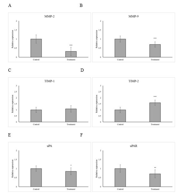
Figure 5. Effect of Ziziphora clinopodioides extract (IC₅₀ concentration, 24 hr) on gene expression of metastasis-related genes in SW742 cells. Relative mRNA levels of (A) MMP-2, (B) MMP-9, (C) TIMP-1, (D) TIMP-2, (E) uPA, and (F) uPAR were measured by Real-time PCR. * p < 0.05, ** p < 0.01, *** p < 0.001 vs. control. (Prepared by Authors, 2026).
Full-Text: (27 Views)
1. Introduction
Colorectal cancer constitutes a major global health burden and the second leading cause of cancer mortality worldwide (1). Despite advances in diagnosis and treatment, metastasis remains the leading cause of CRC-related mortality (2). Approximately 25% of patients present with metastatic disease at diagnosis, and nearly half develop metastases during their illness (3).
Tumor progression toward invasive and metastatic phenotypes is a multifaceted process involving several sequential events, with matrix metalloproteinases (MMPs) and the urokinase-type plasminogen activator (uPA) system serving as key mediators of extracellular matrix degradation and tissue invasion (4). Overexpression of MMP-2, MMP-9, uPA, and uPAR promotes extracellular matrix (ECM) degradation, facilitating tumor spread. Conversely, tissue inhibitors of metalloproteinases (TIMPs) particularly TIMP-1 and TIMP-2 suppress MMP activity and limit metastasis (5).
Natural products have been increasingly studied as potential sources of anticancer compounds due to their multitargeted mechanisms and lower toxicity (6). Ziziphora clinopodioides (family Lamiaceae) has been used widely for various therapeutic purposes in Iranian folk medicine (7). Its essential oil and extracts contain several biologically active components including thymol, carvacrol, 1,8-cineole, limonene, and rosmarinic acid known for antimicrobial, antioxidant, and anticancer effects (8).
Given these properties and the growing interest in plant-derived anticancer agents, this study aimed to evaluate the cytotoxic and antimetastatic potential of Z. clinopodioides extract on colorectal cancer cells.
2. Materials and Methods
2.1 Preparation of Z. clinopodioides extract
The aerial parts of Z. clinopodioides were purchased from a verified medicinal plant supplier (Giah Kala, Tehran, Iran). Following botanical identification and authentication, the collected plant material was air-dried at ambient temperature and subsequently ground into a fine powder. Forty grams of powder were macerated in 300 mL of 70% ethanol (selected for its efficiency in extracting both polar and non-polar phytochemicals) for 72 h on a shaker at room temperature in the dark. After filtration using Whatman No. 1 paper, the extract was subjected to solvent evaporation under controlled conditions at 45 °C for 48 h. The concentrated extract obtained was maintained at −20 °C until use in the experimental procedures.
2.2 Cell culture and treatment
Human SW742 colorectal carcinoma cells were purchased from the Pasteur Institute (Tehran, Iran) and cultured in RPMI-1640 medium supplemented with 10% fetal bovine serum and 1% penicillin–streptomycin. Cells were maintained at 37 °C in a humidified incubator with 5% CO₂. Upon completion of four passages, the cells were subjected to treatment with graded concentrations of the extract (6.25, 12.5, 25, 50, 100, 200, 400, and 800 µg/ml) for viability and cytotoxicity assessments. The IC₅₀ value derived from these experiments was selected for use in subsequent analyses.
2.3 Viability assay
The cells were seeded in 96-well plates (15×103 cells/well) and placed in an incubator for 24 hr to allow the cells to adhere to the bottom of the plate. Then, the culture medium was replaced with the medium containing the extract. The control cells received culture medium. After 24, 48, 72, and 96 hr, 50 μL of MTT solution (5 mg/ml) was added. After 4 hr, the medium was removed and 200 μl of DMSO was added to each well. The plate was subsequently placed on an orbital shaker and mixed for 15 min. Absorbance values were measured at 570 nm, while readings at 650 nm were used for background correction. Cell viability was calculated as the percentage of absorbance in treated cells relative to control cells according to the formula below:
Cell viability (%) = (OD treated / OD control) × 100
IC₅₀ values were estimated using GraphPad Prism 5.0 (USA) based on the dose–response curves generated from the viability data.
2.4 Cytotoxicity assay
The extent of cell membrane damage was determined by analyzing LDH release at 24, 48, 72, and 96 h after treatment. Supernatants were collected and subjected to LDH quantification using a commercially available kit (Sigma, St. Louis, MO, USA) following the manufacturer's guidelines. Absorbance was measured at a wavelength of 490 nm using an ELISA plate reader.
2.5 Cell migration assay
The effect of the extract on cell migration was investigated using a wound-healing (scratch) assay. Cells were plated in 6-well culture plates at a density of 150 × 10³ cells/well and cultured until a uniform monolayer was established. A scratch was created in the cell layer with a sterile pipette tip, after which the wells were gently washed with PBS to eliminate detached cells. The cultures were then maintained in medium containing 2% FBS in the presence or absence of the extract and incubated for 24 h at 37 °C. The migratory capacity of the cells was quantified by monitoring wound closure under a microscope and analyzing the images with T-Scratch software.
2.6 Cell invasion assay
Cell invasion was determined using Transwell chambers (Cell Biolabs, Inc. San Diego, CA, USA) with 6.5 mm polycarbonate filters of 8 μm pore size. 50 μL of Matrigel was pipetted into the upper chamber and incubated at 37 ° C for 30 min to solidify the gel. Cells were seeded at 1×10⁶ cells/mL in 100 μL of the serum-free medium in the upper chamber. Immediately, this chamber was transferred to the lower chamber consisting of 600 μl of medium (2% FBS with and without the extract). After a 24-h incubation period at 37 °C, the upper inserts were processed to remove cells that had not migrated through the membrane. The cells attached to the external surface of the chamber were fixed with 70% methanol for 10 min and stained with 0.2% crystal violet. Following rinsing with distilled water, the samples were dried at ambient temperature and examined under an inverted microscope. The number of migrated cells was quantified by extracting the bound dye with an acetic acid solution and measuring the absorbance at 570 nm.
2.7 Cell adhesion assay
The wells of 6-well culture plates were pre-coated with 0.1% gelatin and dried at 37 °C for 30 min. Subsequently, harvested cells obtained by trypsin/EDTA treatment were suspended in fresh medium in the presence or absence of the extract prior to the adhesion assay. Then, the cells were distributed in the respective wells and incubated at 37 ° C for 24 hr. Finally, the cells were fixed using 70% methanol for 10 min. Then, they were stained with 0.2% crystal violet and washed with distilled water. The cells were air-dried. Then, they were observed under an inverted microscope. The color of the cells was extracted with acetic acid solution and the intensity of the extracted color of each group was measured at a wavelength of 570 nm.
2.8 Gene expression analysis
After treatment, total RNA from cells was extracted using RNXplus solution. The absorbance of each sample was measured at wavelengths 260, 280, and 230, and the ratio of absorbance of 260 to 280 and 260 to 230, as well as RNA concentration, was calculated. The quality of extracted RNA was determined by agarose gel electrophoresis. Considering that RNA cannot be directly used as a template for a Real-time PCR assay, it must first be reverse transcribed into complementary DNA (cDNA). The Ekta Tehiz Teb kit was used to prepare cDNA according to the kit protocol. The resulting cDNA was kept in a freezer at -80 ° C. The specific primers for each gene were designed using Allel ID6 software. GAPDH gene was used as a housekeeping gene to determine the relative expression level of the studied genes. The primer efficiencies were tested and confirmed to be within acceptable limits (90–110%) prior to use (9). The SYBR kit produced by Ben Yakhte Company (Iran) was used for Real-time PCR.
2.9 Statistical analysis
Data are reported as mean values with corresponding standard deviations (mean ± SD). Group differences were analyzed by one-way ANOVA with Tukey’s multiple comparison test, and statistical significance was defined as p < 0.05.
3. Result
3.1 The effect of the extract on cell viability and calculation of IC50
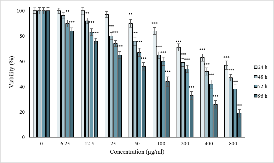
The effects of different concentrations of the extract on cell viability after 24, 48, 72, and 96 h are presented in Figure 1A. After 24 hr, the decrease in cell survival was significant at concentrations of 50, 100, 200, 400, and 800 µg/ml (p<0.05). After 48 hr, it was significant in concentrations of 12.5, 25, 50, 100, 200, 400, and 800 µg/ml (p<0.05) and after 72 and 96 hr, at all concentrations used (p<0.05). The IC50 values of the extract were 760.59 ± 32.45, 395.16 ± 12.85, 241.87 ± 27.77, and 119.05 ± 11.05 for 24, 48, 72, and 96 hr.
3.2 The cytotoxic effect of the extract
After 24 hr, cytotoxicity was significant at concentrations of 100, 200, 400, and 800 µg/ml (p<0.05). After 48 and 72 hr, this effect was significant in concentrations of 25, 50, 100, 200, 400, and 800 μg/ml (p<0.05), and after 96 hr it was significant in concentrations of 12.5, 25, 50, 100, 200, 400 and 800 μg/ml (p<0.05) (Figure 1 B).
3.3 The effect of the extract on cell migration, invasion, and adhesion
Treatment with the extract caused a significant decrease in the ability of cells to migrate and invade (p<0.05) (Figures 2 and Figure 3). It also significantly reduced the adhesion potential of cells (p<0.05) (Figure 4).
A
B
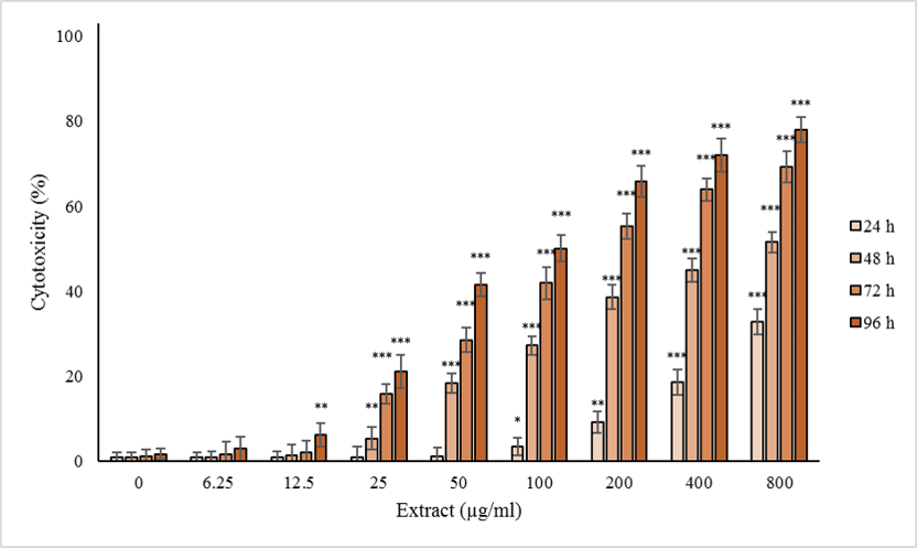
Figure 1. Effect of Ziziphora clinopodioides extract on (A) cell viability and (B) plasma membrane integrity in SW742 colorectal cancer cells. Cells were treated with various concentrations of extract (6.25–800 µg/ml) for 24–96 hr. Cell viability was measured by MTT assay, and plasma membrane integrity was evaluated using the LDH release assay. * p < 0.05, ** p < 0.01, *** p < 0.001 vs. control. (Prepared by Authors, 2026).
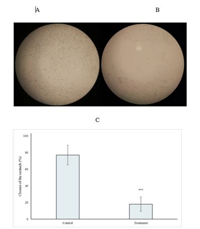
Figure 2. Effect of Ziziphora clinopodioides extract on cell migration ability (scratch assay). (A) Control cells and (B) extract-treated cells after 24 hr. (C) Quantitative analysis of wound closure using T-Scratch software. *** p < 0.001 vs. control. (Prepared by Authors, 2026).
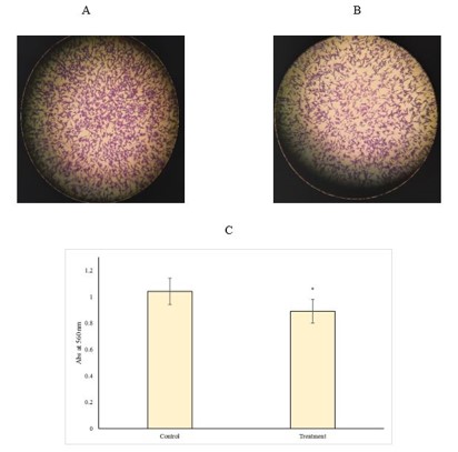
Figure 3. Inhibitory effect of Ziziphora clinopodioides extract on cell invasion. (A) Control cells and (B) extract-treated cells after 24 hr. (C) Quantitative absorbance of crystal-violet-stained invading cells measured at 570 nm. * p < 0.05 vs. control. (Prepared by Authors, 2026).
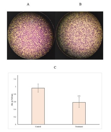
Figure 4. Effect of Ziziphora clinopodioides extract on cell adhesion. (A) Control cells and (B) extract-treated cells after 24 hr. (C) Mean absorbance of attached cells stained with crystal violet at 570 nm. *** p < 0.001 vs. control. (Prepared by Authors, 2026).
3.4 The effect of the extract on gene expression
Quantitative real-time PCR analysis revealed that treatment of CRC cells with the IC₅₀ concentration of the extract for 24 h resulted in a significant reduction in the expression levels of MMP-2 and MMP-9 by 68% and 30%, respectively, compared with the control group (p < 0.05; Figure 5A and B). The expression level of TIMP-1 exhibited a 1.08-fold increase; however, this change was not statistically significant (p < 0.05; Figure 5C). In contrast, TIMP-2 expression was significantly elevated by 1.59-fold following treatment (p < 0.05; Figure 5D). Furthermore, the expression of uPA and uPAR was significantly decreased by 15% and 29%, respectively, relative to untreated cells (p < 0.05; Figures 5E and F).
Quantitative real-time PCR analysis revealed that treatment of CRC cells with the IC₅₀ concentration of the extract for 24 h resulted in a significant reduction in the expression levels of MMP-2 and MMP-9 by 68% and 30%, respectively, compared with the control group (p < 0.05; Figure 5A and B). The expression level of TIMP-1 exhibited a 1.08-fold increase; however, this change was not statistically significant (p < 0.05; Figure 5C). In contrast, TIMP-2 expression was significantly elevated by 1.59-fold following treatment (p < 0.05; Figure 5D). Furthermore, the expression of uPA and uPAR was significantly decreased by 15% and 29%, respectively, relative to untreated cells (p < 0.05; Figures 5E and F).
Figure 5. Effect of Ziziphora clinopodioides extract (IC₅₀ concentration, 24 hr) on gene expression of metastasis-related genes in SW742 cells. Relative mRNA levels of (A) MMP-2, (B) MMP-9, (C) TIMP-1, (D) TIMP-2, (E) uPA, and (F) uPAR were measured by Real-time PCR. * p < 0.05, ** p < 0.01, *** p < 0.001 vs. control. (Prepared by Authors, 2026).
4. Discussion
The present study demonstrated that the 70 % ethanolic extract of Z. clinopodioides significantly inhibited viability, migration, invasion and adhesion of human colorectal cancer (CRC) SW742 cells in a dose- and time-dependent manner. Specifically, the extract reduced IC₅₀ values from ~760 µg/ml at 24 h to ~119 µg/ml at 96 h, and significantly down-regulated expression of metastasis-associated genes (MMP-2, MMP-9, uPA, uPAR) while up-regulating TIMP-2 expression. These findings align with an antimetastatic profile for the extract and provide a mechanistic basis for further investigation.
Our choice of Z. clinopodioides is supported by accumulating phytochemical and pharmacological evidence. The plant belongs to the Lamiaceae family and is rich in flavonoids, phenolic acids and essential oils such as thymol, carvacrol, acacetin, quercetin, apigenin and rhoifolin (8). For example, an LC-MS/MS study identified quinic acid, malic acid, and rhoifolin as major constituents in Z. clinopodioides with antioxidant and enzyme-inhibitory activities (8). Another study on total flavonoids isolated from Z. clinopodioides (ZCF) demonstrated protective effects in oxidative-stress and inflammation models via suppression of VEGF/AKT/NF-κB signaling. Although direct anticancer data on this species are still limited, studies on other Ziziphora spp. have shown cytotoxicity in human cancer cell lines (e.g., Z. tenuior EO in HT-29 colorectal cells) (10). This prior evidence justifies our focus on antimetastatic endpoints in CRC cells. The ability of the extract to down-regulate MMP-2 and MMP-9 is noteworthy because these gelatinases facilitate extracellular matrix (ECM) degradation and tumor invasion. Likewise, the reduction in uPA and its receptor uPAR – key components of the plasminogen activation system – further supports an antimigratory phenotype. Up-regulation of TIMP-2 acts as a counter-regulator of MMPs, reinforcing inhibition of metastatic potential.
From a mechanistic perspective, while our current data are limited to mRNA expression changes, available literature suggests plausible upstream signaling pathways. For example, flavonoids from Z. clinopodioides have been shown to reduce activation of NF-κB and AKT in vascular/inflammatory models (9), and in atherosclerosis models, ZCF suppressed p-PI3K, p-AKT, and p-mTOR expression, while enhancing the autophagy marker LC3 (11). These pathways (PI3K/AKT/mTOR, NF-κB) are well-established regulators of MMPs, uPA/uPAR, and cell motility in cancer. Thus, it is plausible that Z. clinopodioides extract interferes with these signaling cascades in CRC cells, leading to decreased expression of metastasis-promoting genes. Future work should test this by measuring pathway activation (e.g., phospho-AKT, phospho-mTOR, NF-κB nuclear translocation) and evaluating whether specific phytochemicals of the extract bind to target proteins by docking or pull-down assays.
Compared to prior investigations, our study adds new information in several ways. First, while much of the literature on Z. clinopodioides focuses on antioxidant, anti-inflammatory or antimicrobial effects (12), to our knowledge this is among the first studies to examine antimetastatic properties in colorectal cancer cells (migration, invasion, adhesion) with gene expression analysis of MMP/uPA systems. Second, by using a hydroalcoholic extract and standardized cell-based assays, we provide a more physiologically relevant approach than some essential-oil-only studies. The inclusion of gene expression (fold change relative to GAPDH) adds a mechanistic layer. Given the paucity of data on this species in CRC models, the present work therefore represents a novel contribution, although its translational potential remains preliminary.
The growing interest in medicinal plants as potential sources of novel anticancer agents is supported by the diverse bioactive phytochemicals they contain, which can modulate multiple molecular pathways involved in colorectal carcinogenesis and tumor progression. Previous studies have demonstrated that plant-derived compounds can exert antiproliferative, pro-apoptotic, anti-inflammatory, and antimetastatic effects in colorectal cancer models, including the suppression of cellular migration, invasion, adhesion, and angiogenic processes (13–15).
Some limitations should be acknowledged. The extract was not chemically standardized to a defined content of phenolics or major constituents; thus batch-to-batch variability may exist. In vitro findings may not translate in vivo due to bioavailability, metabolism, and pharmacokinetics of the complex extract. Furthermore, while we observed gene expression changes, protein levels, enzyme activities (e.g., gelatine zymography for MMP-2/9), and functional signaling assays were not performed. Finally, the use of a single cell line (SW742) limits generalizability across diverse CRC models. These limitations highlight the need for further work including compound isolation, pathway validation and animal model testing.
To advance the findings reported here, several next steps are recommended. Isolation and characterization of the major bioactive constituents (e.g., acacetin, thymol, carvacrol) should be performed, followed by dose–response studies in multiple CRC cell lines and primary tumor cells. Mechanistic studies should evaluate signaling pathways such as PI3K/AKT/mTOR, NF-κB, MAPK, and WNT/β-catenin, and verify that modulation of MMP/uPA systems is downstream of these pathways. In vivo experiments using CRC metastasis models are necessary to determine efficacy, toxicity, and pharmacokinetics.
Ultimately, while the extract shows potential antimetastatic activity, it should be described as a promising in vitro candidate rather than a therapeutic agent until validated in animal and human studies. Although the data are encouraging, it is critical to temper language regarding therapeutic potential. The present extract should be considered as a preliminary antimetastatic agent pending further validation. Its use in humans cannot be recommended at this stage. Future clinical translation will require rigorous safety profiling, quality-control standardization and demonstration of efficacy in vivo.
5. Conclusion
The hydroalcoholic extract of Z. clinopodioides exhibited cytotoxic and anti-metastatic effects on human colorectal cancer cells in vitro by regulating the expression of metastasis-related genes (MMPs, TIMPs, uPA, and uPAR). These preliminary findings indicate potential anticancer activity that warrants further validation through in vivo and clinical investigations.
6. Declarations
6.1 Acknowledgments
The authors want to thank the Vice-Chancellor for research of Kermanshah University of Medical Sciences for financial support.
6.2 Ethical Considerations
This research project was approved by Kermanshah University of Medical Sciences (IR.KUMS.MED.REC.1401.076 and IR.KUMS.MED.REC.1401.050).
6.3 Authors' Contributions
A. P. designed the experiments, supervised the research, and co-authored the manuscript. C. J, and M. P. performed the cell culture experiments. M. G. analyzed data. K. A. wrote the manuscript. S. S. M. carried out one gene expression analysis of this work. All authors read and approved the final manuscript.
6.4 Conflict of Interest
The authors have no conflicts of interest to declare that are relevant to the content of this article.
6.5 Fund or Financial Support
This study was funded in full by the Vice-Chancellor for Research of Kermanshah University of Medical Sciences, grant number [4010373 and 4010360].
6.6 Using Artificial Intelligence Tools (AI Tools)
Artificial intelligence (AI) tools were used solely to assist with the writing and language editing of this manuscript. AI tools were not involved in data generation, data collection, data analysis, or interpretation of the results. The authors reviewed and revised all AI-assisted content and take full responsibility for the accuracy, integrity, and final content of the manuscript.
The present study demonstrated that the 70 % ethanolic extract of Z. clinopodioides significantly inhibited viability, migration, invasion and adhesion of human colorectal cancer (CRC) SW742 cells in a dose- and time-dependent manner. Specifically, the extract reduced IC₅₀ values from ~760 µg/ml at 24 h to ~119 µg/ml at 96 h, and significantly down-regulated expression of metastasis-associated genes (MMP-2, MMP-9, uPA, uPAR) while up-regulating TIMP-2 expression. These findings align with an antimetastatic profile for the extract and provide a mechanistic basis for further investigation.
Our choice of Z. clinopodioides is supported by accumulating phytochemical and pharmacological evidence. The plant belongs to the Lamiaceae family and is rich in flavonoids, phenolic acids and essential oils such as thymol, carvacrol, acacetin, quercetin, apigenin and rhoifolin (8). For example, an LC-MS/MS study identified quinic acid, malic acid, and rhoifolin as major constituents in Z. clinopodioides with antioxidant and enzyme-inhibitory activities (8). Another study on total flavonoids isolated from Z. clinopodioides (ZCF) demonstrated protective effects in oxidative-stress and inflammation models via suppression of VEGF/AKT/NF-κB signaling. Although direct anticancer data on this species are still limited, studies on other Ziziphora spp. have shown cytotoxicity in human cancer cell lines (e.g., Z. tenuior EO in HT-29 colorectal cells) (10). This prior evidence justifies our focus on antimetastatic endpoints in CRC cells. The ability of the extract to down-regulate MMP-2 and MMP-9 is noteworthy because these gelatinases facilitate extracellular matrix (ECM) degradation and tumor invasion. Likewise, the reduction in uPA and its receptor uPAR – key components of the plasminogen activation system – further supports an antimigratory phenotype. Up-regulation of TIMP-2 acts as a counter-regulator of MMPs, reinforcing inhibition of metastatic potential.
From a mechanistic perspective, while our current data are limited to mRNA expression changes, available literature suggests plausible upstream signaling pathways. For example, flavonoids from Z. clinopodioides have been shown to reduce activation of NF-κB and AKT in vascular/inflammatory models (9), and in atherosclerosis models, ZCF suppressed p-PI3K, p-AKT, and p-mTOR expression, while enhancing the autophagy marker LC3 (11). These pathways (PI3K/AKT/mTOR, NF-κB) are well-established regulators of MMPs, uPA/uPAR, and cell motility in cancer. Thus, it is plausible that Z. clinopodioides extract interferes with these signaling cascades in CRC cells, leading to decreased expression of metastasis-promoting genes. Future work should test this by measuring pathway activation (e.g., phospho-AKT, phospho-mTOR, NF-κB nuclear translocation) and evaluating whether specific phytochemicals of the extract bind to target proteins by docking or pull-down assays.
Compared to prior investigations, our study adds new information in several ways. First, while much of the literature on Z. clinopodioides focuses on antioxidant, anti-inflammatory or antimicrobial effects (12), to our knowledge this is among the first studies to examine antimetastatic properties in colorectal cancer cells (migration, invasion, adhesion) with gene expression analysis of MMP/uPA systems. Second, by using a hydroalcoholic extract and standardized cell-based assays, we provide a more physiologically relevant approach than some essential-oil-only studies. The inclusion of gene expression (fold change relative to GAPDH) adds a mechanistic layer. Given the paucity of data on this species in CRC models, the present work therefore represents a novel contribution, although its translational potential remains preliminary.
The growing interest in medicinal plants as potential sources of novel anticancer agents is supported by the diverse bioactive phytochemicals they contain, which can modulate multiple molecular pathways involved in colorectal carcinogenesis and tumor progression. Previous studies have demonstrated that plant-derived compounds can exert antiproliferative, pro-apoptotic, anti-inflammatory, and antimetastatic effects in colorectal cancer models, including the suppression of cellular migration, invasion, adhesion, and angiogenic processes (13–15).
Some limitations should be acknowledged. The extract was not chemically standardized to a defined content of phenolics or major constituents; thus batch-to-batch variability may exist. In vitro findings may not translate in vivo due to bioavailability, metabolism, and pharmacokinetics of the complex extract. Furthermore, while we observed gene expression changes, protein levels, enzyme activities (e.g., gelatine zymography for MMP-2/9), and functional signaling assays were not performed. Finally, the use of a single cell line (SW742) limits generalizability across diverse CRC models. These limitations highlight the need for further work including compound isolation, pathway validation and animal model testing.
To advance the findings reported here, several next steps are recommended. Isolation and characterization of the major bioactive constituents (e.g., acacetin, thymol, carvacrol) should be performed, followed by dose–response studies in multiple CRC cell lines and primary tumor cells. Mechanistic studies should evaluate signaling pathways such as PI3K/AKT/mTOR, NF-κB, MAPK, and WNT/β-catenin, and verify that modulation of MMP/uPA systems is downstream of these pathways. In vivo experiments using CRC metastasis models are necessary to determine efficacy, toxicity, and pharmacokinetics.
Ultimately, while the extract shows potential antimetastatic activity, it should be described as a promising in vitro candidate rather than a therapeutic agent until validated in animal and human studies. Although the data are encouraging, it is critical to temper language regarding therapeutic potential. The present extract should be considered as a preliminary antimetastatic agent pending further validation. Its use in humans cannot be recommended at this stage. Future clinical translation will require rigorous safety profiling, quality-control standardization and demonstration of efficacy in vivo.
5. Conclusion
The hydroalcoholic extract of Z. clinopodioides exhibited cytotoxic and anti-metastatic effects on human colorectal cancer cells in vitro by regulating the expression of metastasis-related genes (MMPs, TIMPs, uPA, and uPAR). These preliminary findings indicate potential anticancer activity that warrants further validation through in vivo and clinical investigations.
6. Declarations
6.1 Acknowledgments
The authors want to thank the Vice-Chancellor for research of Kermanshah University of Medical Sciences for financial support.
6.2 Ethical Considerations
This research project was approved by Kermanshah University of Medical Sciences (IR.KUMS.MED.REC.1401.076 and IR.KUMS.MED.REC.1401.050).
6.3 Authors' Contributions
A. P. designed the experiments, supervised the research, and co-authored the manuscript. C. J, and M. P. performed the cell culture experiments. M. G. analyzed data. K. A. wrote the manuscript. S. S. M. carried out one gene expression analysis of this work. All authors read and approved the final manuscript.
6.4 Conflict of Interest
The authors have no conflicts of interest to declare that are relevant to the content of this article.
6.5 Fund or Financial Support
This study was funded in full by the Vice-Chancellor for Research of Kermanshah University of Medical Sciences, grant number [4010373 and 4010360].
6.6 Using Artificial Intelligence Tools (AI Tools)
Artificial intelligence (AI) tools were used solely to assist with the writing and language editing of this manuscript. AI tools were not involved in data generation, data collection, data analysis, or interpretation of the results. The authors reviewed and revised all AI-assisted content and take full responsibility for the accuracy, integrity, and final content of the manuscript.
Type of Study: Original Research Article |
Subject:
Life Science
Received: 2026/05/3 | Accepted: 2026/08/16 | Published: 2026/08/4
Received: 2026/05/3 | Accepted: 2026/08/16 | Published: 2026/08/4
References
1. Rawla P, Sunkara T, Barsouk A. Epidemiology of colorectal cancer: incidence, mortality, survival, and risk factors. Prz Gastroenterol. 2019;14(2):89-103. [DOI:10.5114/pg.2018.81072] [PMID] [PMCID]
2. Shin AE, Giancotti FG, Rustgi AK. Metastatic colorectal cancer: mechanisms and emerging therapeutics. Trends Pharmacol Sci. 2023;44(4):222-36. [DOI:10.1016/j.tips.2023.01.003] [PMID] [PMCID]
3. Van Cutsem E, De Gramont A, Henning G, Rougier P, Bonnetain F, Seufferlein T. Improving outcomes in patients with CRC: the role of patient reported outcomes-an ESDO report. Cancers (Basel). 2017;9(6):59. [DOI:10.3390/cancers9060059] [PMID] [PMCID]
4. Baker EA, Leaper DJ. The plasminogen activator and matrix metalloproteinase systems in colorectal cancer: relationship to tumour pathology. Eur J Cancer. 2003;39(7):981-8. [DOI:10.1016/S0959-8049(03)00065-0] [PMID]
5. Fan SQ, Wei QY, Li MR, Zhang LQ, Liang QC. Expression and clinical significance of MMP-2, MMP-9, TIMP-1, and TIMP-2 in breast carcinoma. Ai Zheng. 2003;22(9):968-73.
6. Asma ST, Acaroz U, Imre K, Morar A, Shah SRA, Hussain SZ, et al. Natural products/bioactive compounds as a source of anticancer drugs. Cancers (Basel). 2022;14(24):6203 [DOI:10.3390/cancers14246203] [PMID] [PMCID]
7. Mohammadifard F, Alimohammadi S. Chemical composition and role of opioidergic system in antinociceptive effect of Ziziphora clinopodioides essential oil. Basic Clin Neurosci. 2018;9(5):357-66. [DOI:10.32598/bcn.9.5.357] [PMID] [PMCID]
8. Mazarei Z. Chemical composition and biological activity of Ziziphora clinopodioides essential oil: a review. Jundishapur J Nat Pharm Prod. 2023;18(4):e137776. [DOI:10.5812/jjnpp-137776]
9. Movahhed M, Pazhouhi M, Ghaleh HEG, Kondori BJ. Anti-metastatic effect of taraxasterol on prostate cancer cell lines. Res Pharm Sci. 2023;18(4):439-48. [DOI:10.4103/1735-5362.378090] [PMID] [PMCID]
10. Zhaparkulova K, Karaubayeva A, Sakipova Z, Biernasiuk A, Gaweł-Bęben K, Laskowski T, et al. Multidirectional characterization of phytochemical profile and health-promoting effects of Ziziphora bungeana Juz. extracts. Molecules. 2022;27(24):8994. [DOI:10.3390/molecules27248994] [PMID] [PMCID]
11. Ma XY, Zhao HR, Qiao HL, Zeng YC, Zhang XM. Mechanism of total flavonoids of Ziziphora clinopodioides in improving atherosclerosis by regulating PI3K/Akt/mTOR pathway. Zhongguo Zhong Yao Za Zhi. 2023;48:465-71.
12. Omer Qader K, Malik Al-Saadi SAA, Hiwa Arif H, Al-Fekaiki DF. Antibacterial and antioxidant activity of Ziziphora clinopodioides Lam. (Lamiaceae) essential oil. Arch Razi Inst. 2023;78(1):205-11.
13. Li YH, Niu YB, Sun Y, Zhang F, Liu CX, Fan L, Mei QB. Role of phytochemicals in colorectal cancer prevention. World J Gastroenterol. 2015;21(31):9262-72. [DOI:10.3748/wjg.v21.i31.9262] [PMID] [PMCID]
14. Benarba B, Pandiella A. Colorectal cancer and medicinal plants: Principle findings from recent studies. Biomed Pharmacother. 2018;107:408-23. [DOI:10.1016/j.biopha.2018.08.006] [PMID]
15. Aiello P, Sharghi M, Mansourkhani SM, Ardekania R, Jouybari L, Lamashti AM, et al. Medicinal plants in the prevention and treatment of colon cancer. Evid Based Complement Alternat Med. 2019;2019:2075614. [DOI:10.1155/2019/2075614] [PMID] [PMCID]
Send email to the article author
| Rights and permissions | |
 |
This work is licensed under a Creative Commons Attribution-NonCommercial 4.0 International License. |
Copyright Policy
Journal of Advances in Medical and Biomedical Research by Zanjan University of Medical Sciences is licensed under CC BY-NC 4.0