
Sun, Sep 20, 2026
[Archive]
Volume 34, Issue 2 (March & April 2026)
J Adv Med Biomed Res 2026, 34(2): 110-119 |
Back to browse issues page



Download citation:
BibTeX | RIS | EndNote | Medlars | ProCite | Reference Manager | RefWorks
Send citation to:


BibTeX | RIS | EndNote | Medlars | ProCite | Reference Manager | RefWorks
Send citation to:
Rahmani S, Mohammadi M, Soleimani B, Mansouri K, Maleki A. Design of an RT-qPCR Assay for Detecting PML-RARA Fusion Gene in Acute Promyelocytic Leukemia Patients. J Adv Med Biomed Res 2026; 34 (2) :110-119
URL: http://journal.zums.ac.ir/article-1-7920-en.html
URL: http://journal.zums.ac.ir/article-1-7920-en.html


1- Department of Medical Laboratory Sciences, School of Paramedical, Kermanshah University of Medical Sciences, Kermanshah, Iran
2- Department of Hematology and Blood Banking, School of Allied Medical Sciences, Shahid Beheshti University of Medical Sciences, Tehran, Iran
3- Medical Biology Research Center, Health Technology Institute, Kermanshah University of Medical Sciences, Kermanshah, Iran
4- Medical Biology Research Center, Health Technology Institute, Kermanshah University of Medical Sciences
5- Department of Molecular Medicine, School of Medicine, Kermanshah University of Medical Sciences, Kermanshah, Iran ,maleki.hem@gmail.com
2- Department of Hematology and Blood Banking, School of Allied Medical Sciences, Shahid Beheshti University of Medical Sciences, Tehran, Iran
3- Medical Biology Research Center, Health Technology Institute, Kermanshah University of Medical Sciences, Kermanshah, Iran
4- Medical Biology Research Center, Health Technology Institute, Kermanshah University of Medical Sciences
5- Department of Molecular Medicine, School of Medicine, Kermanshah University of Medical Sciences, Kermanshah, Iran ,
Keywords: Key words: Acute Promyelocytic Leukemia (APL), PML-RARA fusion, bcr1 variant, Translocation t (15, 17)
Full-Text [PDF 540 kb]
(159 Downloads)
| Abstract (HTML) (750 Views)
Full-Text: (18 Views)
ABSTRACT
Background & Objective: Acute promyelocytic leukemia (APL) is a subtype of acute myeloid leukemia characterized by the t(15;17) translocation, generating the PML-RARA fusion gene. This fusion occurs in three primary variants: bcr1, bcr2, and bcr3. In Iran, bcr1 is the most prevalent (~73%), bcr3 accounts for ~27%, and bcr2 is virtually undetected. Given this distribution, developing a locally optimized method for detecting bcr1 is essential for accurate and rapid identification of this fusion and crucial for monitoring patient response.
Materials & Methods: A synthetic 130-bp PML-RARA bcr1 fragment was cloned into pUC57, and insertion was analyzed by digestion with XbaI and HindIII and agarose gel electrophoresis. RNA from APL patient samples was reverse-transcribed into cDNA, serving as a template for reverse-transcription quantitative polymerase chain reaction (RT-qPCR) with PML-RARA-specific primers and β-actin as the reference. Each run included APL samples, plasmid-based positive controls, healthy negative controls, and no-template controls. Amplification and melt-curve analysis confirmed assay specificity and reproducibility.
Results: The recombinant pUC57-PML-RARA plasmid was verified by XbaI/HindIII digestion. RT-qPCR showed specific amplification of PML-RARA in plasmid controls (Ct 15-20) and APL patient samples (Ct 22-26), with no amplification in negative controls. Melt curve analysis showed single, sharp peaks, confirming specificity, and agarose gel electrophoresis verified the expected product sizes. Relative quantification indicated approximately 26-fold higher expression in plasmid controls compared to patient samples, with high reproducibility at a 1:20 dilution.
Conclusion: This preliminary assessment shows sufficient specificity and reliability for detecting the PML-RARA bcr1 variant, providing a foundation for further validation in the future.
Keywords: Acute Promyelocytic Leukemia (APL), PML-RARA fusion, bcr1 variant, Translocation t
Acute promyelocytic leukemia is a clinically and genetically distinct subtype of acute myeloid leukemia (AML), defined by the reciprocal translocation t (15;17) (q24; q21), which results in the PML-RARA fusion gene. The resultant fusion protein disrupts normal myeloid differentiation, leading to an accumulation of promyelocytes and severe clinical manifestations, including coagulopathy (1-4).
Molecularly, PML-RARA exists in three principal variants: bcr1, bcr2, and bcr3 (5,6). While their global distribution is relatively balanced, epidemiological studies in Iran reveal a distinct pattern: bcr1 predominates (~73%), bcr3 is observed in ~27% of cases, and bcr2 has not been reported.
This epidemiological profile underscores the importance of designing diagnostic methods that specifically target the bcr1 variant for the Iranian population (7).
Targeted therapies, particularly all-trans retinoic acid (ATRA) and arsenic trioxide (ATO), have transformed APL treatment by promoting differentiation of leukemic promyelocytes via the PML-RARA oncoprotein. However, their clinical success depends on timely and accurate detection of the fusion transcript, highlighting the need for rapid, sensitive, and cost-effective molecular diagnostics. (8,9).
Conventional cytogenetic techniques, such as karyotyping and fluorescence in situ hybridization (FISH), provide valuable insights but are often limited by low sensitivity, prolonged turnaround times, and reliance on specialized instrumentation (10,11).
Commercial reverse-transcription quantitative polymerase chain reaction (RT-qPCR) kits enable detection of multiple variants but may face challenges including high cost, probe dependency, and validation requirement across different populations (12-14).
In contrast, a targeted RT-qPCR approach employing variant-specific primers and melt curve analysis offers a rapid, accessible, and reliable strategy for detecting predominant variants such as bcr1 without the need for expensive probes (15-17).
Incorporating plasmid-based positive controls further ensures assay accuracy and reproducibility (14).
The present study aimed to develop and preliminarily validate a locally optimized RT-qPCR method for the sensitive and specific detection of the PML-RARA bcr1 fusion in Iranian APL patients, providing a foundation for population-specific molecular diagnostics and future assay validation.
2. Materials and Methods
2.1 Sample Collection
A total of 20 peripheral blood samples were collected at Shariati Hospital, Tehran. Ten samples were obtained from patients diagnosed with APL based on clinical, morphological, and molecular criteria, and ten samples from healthy individuals served as negative controls. All samples were collected in EDTA tubes with RNA-stabilizing buffer (RNA later) and transported on dry ice to the Molecular Biology Research Laboratory for further processing.
All participants provided written informed consent, and the study protocol was approved by the Ethics Committee of Kermanshah University of Medical Sciences.
2.2 RNA extraction and quality assessment
Total RNA was extracted from peripheral blood leukocytes using the Buffy coat layer. Briefly, 5 mL of EDTA-anticoagulated blood was centrifuged at 2,500 g for 10 minutes at room temperature to separate plasma, and the Buffy coat was collected as a source of nucleated cells.
Red blood cells were removed by incubation with an RBC lysis buffer for 5 minutes at room temperature, followed by centrifugation at 300 g; this step was repeated once.
The remaining cell pellet was lysed in TRIzol reagent, followed by chloroform phase separation and isopropanol-mediated RNA precipitation. RNA pellets were washed with 75% ethanol, air-dried, and resuspended in RNase-free water. RNA quantity and purity were assessed using a NanoDrop spectrophotometer, and integrity was evaluated by 1 % agarose gel electrophoresis. Extracted RNA was stored at -80 °C until further use.
2.3 cDNA Synthesis
Complementary DNA (cDNA) was synthesized from DNase I-treated total RNA using a commercial cDNA synthesis kit (Smobio, Taiwan). Briefly, 7 µL of RNA was combined with 1 µL dNTPs, 1 µL random hexamers, and 1 µL oligo(dT) primers in a 0.2 mL tube, followed by denaturation at 70 °C for 5 min and quick chilling on ice. The reverse transcription reaction was carried out by adding 4 µL buffer, 1 µL RNase inhibitor, and 1 µL reverse transcriptase, with incubation at 25 °C for 10 min, 50 °C for 50 min, and 85 °C for 5 min to inactivate the enzyme. The resulting cDNA was stored at -20 °C for subsequent gene expression analysis using RT-qPCR.
2.4 Primer Design for Target Genes
To evaluate the relative expression of PML-RARA and the reference gene β-actin, specific primers were designed based on full-length gene sequences retrieved from the NCBI database.
Primers were developed using Primer-BLAST, adhering to standard criteria: 18-25 nucleotides in length, melting temperatures between 58-62 °C, GC content of 40-60%, and avoidance of secondary structures such as hairpins or dimers.
Amplified product sizes were kept below 200 bp to ensure optimal qPCR efficiency. The final primer sequences were as follows: for PML-RARA, forward 5′-CGACCCCAACGCTAAGACAC-3′ and reverse 5′-GCACGTAGCCATATTTGCCTT-3′, producing a 130 bp fragment; for β-actin, forward 5′-TGACCCAGCCATGTTTGAGA-3′ and reverse 5′-CTCGTAGATGGCACAGTGTGG-3′, producing a 156 bp fragment.
2.5 Plasmid construction
The synthetic PML-RARA sequence was cloned into the pUC57 plasmid between the XbaI and HindIII restriction sites and constructed synthetically by GENERAY (Taiwan).
2.6 Preparation of Competent E. coli DH5α
For plasmid amplification, E. coli DH5α was used as the host strain. Competent cells were prepared following standard protocols with minor modifications. Briefly, an overnight culture of DH5α in LB or SOC medium without antibiotics was diluted 1:20-1:200 and grown at 37 °C with shaking until reaching mid-log phase (OD600 ≈ 0.6-0.8). Cells were chilled on ice, harvested by centrifugation, and washed with cold calcium chloride solution. The final cell pellet was resuspended in a mixture of glycerol and 100 mM calcium chloride, aliquoted, and stored at -70 °C until use.
2.7 Bacterial Transformation
Approximately 100 ng of purified DNA was then gently mixed with competent E. coli DH5α cells and incubated on ice for 30 s. The cells underwent heat-shock at 42 °C for 90 s, followed by a 90 s incubation on ice, to facilitate DNA uptake. Subsequently, 800 µL of LB medium without antibiotic was added, and the cultures were incubated at 37 °C with gentle shaking for 2 h to allow expression of the antibiotic resistance gene. Finally, the transformed cells were plated on LB agar containing 30 µg/mL ampicillin and incubated overnight at 37 °C, while untransformed cells were plated as a negative control.
2.8 Plasmid Extraction and quality assessment
Approximately 5 h after inoculation, plasmid DNA was extracted from selected E. coli DH5α colonies harboring pUC57-PML-RARA using the Mini Plus Plasmid DNA Extraction System (Viogene, Cat. No.: GF2001), following the manufacturer’s instructions. Bacterial cells were harvested by centrifugation, and the pellet was resuspended in the provided buffers. Lysis, neutralization, and washing steps were performed according to the kit protocol. The purified plasmid DNA was finally resuspended in DNase/RNase-free water, and its quality and quantity were assessed by agarose gel electrophoresis and Nanodrop spectrophotometry.
2.9 Enzyme digestion of plasmid
To confirm the presence and correct insertion of the PML-RARA, plasmids were digested with XbaI and HindIII restriction enzymes. Digestion reactions were performed under standard conditions, and the resulting fragments were separated by 1% agarose gel electrophoresis. Clones producing the expected 130 bp fragment were considered positive.
2.10 Real time quantitative PCR (RT-qPCR)
Quantitative assessment of PML-RARA expression was performed using RT-qPCR with β-Actin as an internal reference gene. SYBR Green was employed as the fluorescent dye, and gene-specific primers were used. Reactions were prepared in 0.2 mL PCR tubes with a final volume of 20 µL, containing cDNA, primers, master mix, and nuclease-free water. All sample preparation steps were conducted on ice to prevent degradation. Samples were briefly centrifuged and transferred to the qPCR instrument.
To optimize reaction conditions, a plasmid positive control was tested at five dilutions (1:1, 1:5, 1:10, 1:20, and 1:50), with the 1:20 dilution producing the most consistent amplification profile with minimal noise. Relative gene expression was calculated using the 2-ΔΔCt method, where ΔCt represents the difference between target and reference gene Ct values, ΔΔCt is the difference between treated and control, and relative fold change=2-ΔΔCt. This approach allowed accurate quantification of PML-RARA expression relative to the internal reference.
2.11 Statistical analysis
Quantitative data from RT-qPCR, including Ct values and calculated fold changes, were analyzed using SPSS version 22. Mean ± standard deviation (SD) values were calculated for each group, and relative expression differences between plasmid and patient samples were assessed.
3. Result
3.1 Verification of PML-RARA Cloning
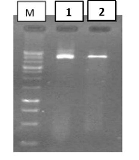
Digestion of the recombinant pUC57-PML-RARA plasmid with XbaI and HindIII yielded the expected fragment sizes. Distinct and well-defined bands were observed on the agarose gel (as shown in Figure 1), confirming the correct insertion of the PML-RARA sequenc.
As depicted in Figure 2, plasmid controls showed amplification within a Ct range of 15-20, with uniform and overlapping curves, indicating high primer efficiency and reaction sensitivity.
Patient samples amplified within a Ct range of 22-26, consistent with clinical specimens. No signal was detected for PML-RARA in healthy control samples (negative controls), confirming the assay’s specificity.
The β-actin gene showed normal expression across all samples, with no significant difference between patients and controls, validating sample quality and extraction efficiency.
Negative RT-PCR controls (without cDNA) showed no amplification, confirming absence of contamination and proper experimental conditions.
3.3 Real-time PCR Melting curves
Melting curve analysis of all plasmid and patient samples for both PML-RARA and β-actin exhibited sharp, single-peak profiles with minimal background noise. The plasmid peak displayed a characteristic amplitude and shape, indicating high reaction specificity and the absence of non-specific amplification or primer-dimer formation (Figure 3).
3.4 Agarose Gel Analysis of RT-qPCR Amplification Products
As shown in Figure 4, RT-qPCR products were analyzed on a 2% agarose gel, showing sharp, single bands at the expected sizes: 156 bp for β‑actin and 130 bp for PML-RARA. No bands were detected in negative controls, confirming high specificity and absence of contamination. These results validate the accurate and specific amplification of the target genes.
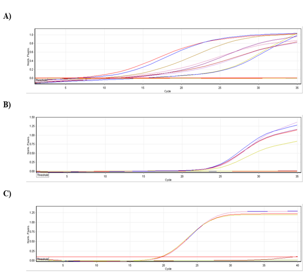
Figure 2. RT-qPCR Amplification plots for PML-RARA. (A) β-Actin as the internal reference gene, (B) patient samples with acute promyelocytic leukemia, and (C) pUC57-PML-RARA plasmid as the positive control. Lower Ct values indicate higher expression levels of the target gene. (Prepared by Authors, 2026).
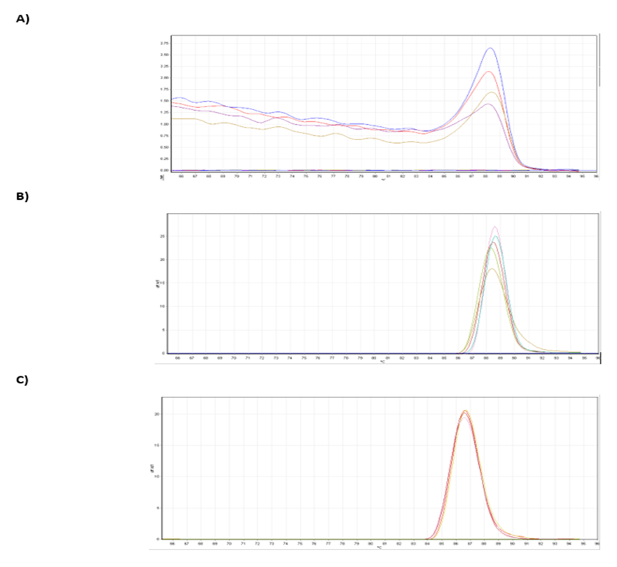
Figure 3. Melt curve analysis of RT-qPCR reactions for PML-RARA: (A) β-actin as the internal reference gene, (B) patient samples with acute promyelocytic leukemia, and (C) plasmid containing PML-RARA as the kit positive control. The presence of distinct single peaks indicates high amplification specificity and the absence of non-specific products or primer-dimer formation. (Prepared by Authors, 2026).
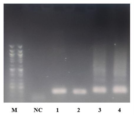
Figure 4. Agarose gel (2%) analysis of RT-qPCR products. Lane M: 50 bp DNA ladder; NC: negative control (no amplification); Lanes 1-2: PML-RARA, 130 bp; Lanes 3-4: β‑actin, 156 bp. All bands are sharp and single, confirming specific amplification. (Prepared by Authors, 2026).
In response to the increasing demand for locally developed diagnostic assays for APL, we report the successful design and preliminary evaluation of a RT-qPCR assay employing a recombinant plasmid positive control harboring the bcr1 variant of the PML-RARA gene. Plasmid-based controls offer distinct advantages, including high target copy number, long-term stability, reproducibility, and scalability for routine laboratory use. The assay demonstrated robust performance, evidenced by consistent Ct values across ten consecutive runs, well-defined single-peak melt curves, and clear discrimination between positive control and patient samples. While this initial design serves as a proof-of-concept, it is limited to detection of the bcr1 variant, with other clinically relevant variants (bcr2, bcr3) yet to be incorporated. Long-term stability and comprehensive clinical validation remain to be established.
Accurate gene insertion into the pUC57 vector was confirmed via double digestion and agarose gel electrophoresis, underscoring the reliability of the plasmid construct. Collectively, these results provide a strong foundation for the development of a fully validated, cost-effective, and locally produced diagnostic platform, with future studies aimed at broadening variant coverage, evaluating plasmid stability over extended periods, and performing multi-center clinical validation.
6. Declarations
6.1 Acknowledgments
We would like to thank the staff of the medical Biology Research center, Kermanshah University of medical sciences, Kermanshah, IR Iran.
6.2 Ethical Considerations
The study protocol was approved by the Ethics Committee of Kermanshah University of Medical Sciences (Ethical code: IR.KUMS.REC.1403.427)
6.3 Authors' Contributions
Ali Maleki and Mohammad Hossein Mohammadi designed and supervised the study. Bijan Soleymani and Kamran Mansouri provided guidance and advisory support. Shahla Rahmani conducted the experiments, collected and analyzed the data, and drafted the manuscript. Ali Maleki critically revised and finalized the manuscript. All authors have read and approved the final manuscript.
6.4 Conflict of Interest
None declared
6.5 Fund or Financial Support
This study was financially supported by Kermanshah University of Medical Sciences (thesis number: 4030799)
6.6 Using Artificial Intelligence Tools (AI Tools)
The authors didn’t use Artificial intelligence tools for writing this manuscript.
Background & Objective: Acute promyelocytic leukemia (APL) is a subtype of acute myeloid leukemia characterized by the t(15;17) translocation, generating the PML-RARA fusion gene. This fusion occurs in three primary variants: bcr1, bcr2, and bcr3. In Iran, bcr1 is the most prevalent (~73%), bcr3 accounts for ~27%, and bcr2 is virtually undetected. Given this distribution, developing a locally optimized method for detecting bcr1 is essential for accurate and rapid identification of this fusion and crucial for monitoring patient response.
Materials & Methods: A synthetic 130-bp PML-RARA bcr1 fragment was cloned into pUC57, and insertion was analyzed by digestion with XbaI and HindIII and agarose gel electrophoresis. RNA from APL patient samples was reverse-transcribed into cDNA, serving as a template for reverse-transcription quantitative polymerase chain reaction (RT-qPCR) with PML-RARA-specific primers and β-actin as the reference. Each run included APL samples, plasmid-based positive controls, healthy negative controls, and no-template controls. Amplification and melt-curve analysis confirmed assay specificity and reproducibility.
Results: The recombinant pUC57-PML-RARA plasmid was verified by XbaI/HindIII digestion. RT-qPCR showed specific amplification of PML-RARA in plasmid controls (Ct 15-20) and APL patient samples (Ct 22-26), with no amplification in negative controls. Melt curve analysis showed single, sharp peaks, confirming specificity, and agarose gel electrophoresis verified the expected product sizes. Relative quantification indicated approximately 26-fold higher expression in plasmid controls compared to patient samples, with high reproducibility at a 1:20 dilution.
Conclusion: This preliminary assessment shows sufficient specificity and reliability for detecting the PML-RARA bcr1 variant, providing a foundation for further validation in the future.
Keywords: Acute Promyelocytic Leukemia (APL), PML-RARA fusion, bcr1 variant, Translocation t
1. Introduction
Molecularly, PML-RARA exists in three principal variants: bcr1, bcr2, and bcr3 (5,6). While their global distribution is relatively balanced, epidemiological studies in Iran reveal a distinct pattern: bcr1 predominates (~73%), bcr3 is observed in ~27% of cases, and bcr2 has not been reported.
This epidemiological profile underscores the importance of designing diagnostic methods that specifically target the bcr1 variant for the Iranian population (7).
Targeted therapies, particularly all-trans retinoic acid (ATRA) and arsenic trioxide (ATO), have transformed APL treatment by promoting differentiation of leukemic promyelocytes via the PML-RARA oncoprotein. However, their clinical success depends on timely and accurate detection of the fusion transcript, highlighting the need for rapid, sensitive, and cost-effective molecular diagnostics. (8,9).
Conventional cytogenetic techniques, such as karyotyping and fluorescence in situ hybridization (FISH), provide valuable insights but are often limited by low sensitivity, prolonged turnaround times, and reliance on specialized instrumentation (10,11).
Commercial reverse-transcription quantitative polymerase chain reaction (RT-qPCR) kits enable detection of multiple variants but may face challenges including high cost, probe dependency, and validation requirement across different populations (12-14).
In contrast, a targeted RT-qPCR approach employing variant-specific primers and melt curve analysis offers a rapid, accessible, and reliable strategy for detecting predominant variants such as bcr1 without the need for expensive probes (15-17).
Incorporating plasmid-based positive controls further ensures assay accuracy and reproducibility (14).
The present study aimed to develop and preliminarily validate a locally optimized RT-qPCR method for the sensitive and specific detection of the PML-RARA bcr1 fusion in Iranian APL patients, providing a foundation for population-specific molecular diagnostics and future assay validation.
2. Materials and Methods
2.1 Sample Collection
A total of 20 peripheral blood samples were collected at Shariati Hospital, Tehran. Ten samples were obtained from patients diagnosed with APL based on clinical, morphological, and molecular criteria, and ten samples from healthy individuals served as negative controls. All samples were collected in EDTA tubes with RNA-stabilizing buffer (RNA later) and transported on dry ice to the Molecular Biology Research Laboratory for further processing.
All participants provided written informed consent, and the study protocol was approved by the Ethics Committee of Kermanshah University of Medical Sciences.
2.2 RNA extraction and quality assessment
Total RNA was extracted from peripheral blood leukocytes using the Buffy coat layer. Briefly, 5 mL of EDTA-anticoagulated blood was centrifuged at 2,500 g for 10 minutes at room temperature to separate plasma, and the Buffy coat was collected as a source of nucleated cells.
Red blood cells were removed by incubation with an RBC lysis buffer for 5 minutes at room temperature, followed by centrifugation at 300 g; this step was repeated once.
The remaining cell pellet was lysed in TRIzol reagent, followed by chloroform phase separation and isopropanol-mediated RNA precipitation. RNA pellets were washed with 75% ethanol, air-dried, and resuspended in RNase-free water. RNA quantity and purity were assessed using a NanoDrop spectrophotometer, and integrity was evaluated by 1 % agarose gel electrophoresis. Extracted RNA was stored at -80 °C until further use.
2.3 cDNA Synthesis
Complementary DNA (cDNA) was synthesized from DNase I-treated total RNA using a commercial cDNA synthesis kit (Smobio, Taiwan). Briefly, 7 µL of RNA was combined with 1 µL dNTPs, 1 µL random hexamers, and 1 µL oligo(dT) primers in a 0.2 mL tube, followed by denaturation at 70 °C for 5 min and quick chilling on ice. The reverse transcription reaction was carried out by adding 4 µL buffer, 1 µL RNase inhibitor, and 1 µL reverse transcriptase, with incubation at 25 °C for 10 min, 50 °C for 50 min, and 85 °C for 5 min to inactivate the enzyme. The resulting cDNA was stored at -20 °C for subsequent gene expression analysis using RT-qPCR.
2.4 Primer Design for Target Genes
To evaluate the relative expression of PML-RARA and the reference gene β-actin, specific primers were designed based on full-length gene sequences retrieved from the NCBI database.
Primers were developed using Primer-BLAST, adhering to standard criteria: 18-25 nucleotides in length, melting temperatures between 58-62 °C, GC content of 40-60%, and avoidance of secondary structures such as hairpins or dimers.
Amplified product sizes were kept below 200 bp to ensure optimal qPCR efficiency. The final primer sequences were as follows: for PML-RARA, forward 5′-CGACCCCAACGCTAAGACAC-3′ and reverse 5′-GCACGTAGCCATATTTGCCTT-3′, producing a 130 bp fragment; for β-actin, forward 5′-TGACCCAGCCATGTTTGAGA-3′ and reverse 5′-CTCGTAGATGGCACAGTGTGG-3′, producing a 156 bp fragment.
2.5 Plasmid construction
The synthetic PML-RARA sequence was cloned into the pUC57 plasmid between the XbaI and HindIII restriction sites and constructed synthetically by GENERAY (Taiwan).
2.6 Preparation of Competent E. coli DH5α
For plasmid amplification, E. coli DH5α was used as the host strain. Competent cells were prepared following standard protocols with minor modifications. Briefly, an overnight culture of DH5α in LB or SOC medium without antibiotics was diluted 1:20-1:200 and grown at 37 °C with shaking until reaching mid-log phase (OD600 ≈ 0.6-0.8). Cells were chilled on ice, harvested by centrifugation, and washed with cold calcium chloride solution. The final cell pellet was resuspended in a mixture of glycerol and 100 mM calcium chloride, aliquoted, and stored at -70 °C until use.
2.7 Bacterial Transformation
Approximately 100 ng of purified DNA was then gently mixed with competent E. coli DH5α cells and incubated on ice for 30 s. The cells underwent heat-shock at 42 °C for 90 s, followed by a 90 s incubation on ice, to facilitate DNA uptake. Subsequently, 800 µL of LB medium without antibiotic was added, and the cultures were incubated at 37 °C with gentle shaking for 2 h to allow expression of the antibiotic resistance gene. Finally, the transformed cells were plated on LB agar containing 30 µg/mL ampicillin and incubated overnight at 37 °C, while untransformed cells were plated as a negative control.
2.8 Plasmid Extraction and quality assessment
Approximately 5 h after inoculation, plasmid DNA was extracted from selected E. coli DH5α colonies harboring pUC57-PML-RARA using the Mini Plus Plasmid DNA Extraction System (Viogene, Cat. No.: GF2001), following the manufacturer’s instructions. Bacterial cells were harvested by centrifugation, and the pellet was resuspended in the provided buffers. Lysis, neutralization, and washing steps were performed according to the kit protocol. The purified plasmid DNA was finally resuspended in DNase/RNase-free water, and its quality and quantity were assessed by agarose gel electrophoresis and Nanodrop spectrophotometry.
2.9 Enzyme digestion of plasmid
To confirm the presence and correct insertion of the PML-RARA, plasmids were digested with XbaI and HindIII restriction enzymes. Digestion reactions were performed under standard conditions, and the resulting fragments were separated by 1% agarose gel electrophoresis. Clones producing the expected 130 bp fragment were considered positive.
2.10 Real time quantitative PCR (RT-qPCR)
Quantitative assessment of PML-RARA expression was performed using RT-qPCR with β-Actin as an internal reference gene. SYBR Green was employed as the fluorescent dye, and gene-specific primers were used. Reactions were prepared in 0.2 mL PCR tubes with a final volume of 20 µL, containing cDNA, primers, master mix, and nuclease-free water. All sample preparation steps were conducted on ice to prevent degradation. Samples were briefly centrifuged and transferred to the qPCR instrument.
To optimize reaction conditions, a plasmid positive control was tested at five dilutions (1:1, 1:5, 1:10, 1:20, and 1:50), with the 1:20 dilution producing the most consistent amplification profile with minimal noise. Relative gene expression was calculated using the 2-ΔΔCt method, where ΔCt represents the difference between target and reference gene Ct values, ΔΔCt is the difference between treated and control, and relative fold change=2-ΔΔCt. This approach allowed accurate quantification of PML-RARA expression relative to the internal reference.
2.11 Statistical analysis
Quantitative data from RT-qPCR, including Ct values and calculated fold changes, were analyzed using SPSS version 22. Mean ± standard deviation (SD) values were calculated for each group, and relative expression differences between plasmid and patient samples were assessed.
3. Result
3.1 Verification of PML-RARA Cloning
Digestion of the recombinant pUC57-PML-RARA plasmid with XbaI and HindIII yielded the expected fragment sizes. Distinct and well-defined bands were observed on the agarose gel (as shown in Figure 1), confirming the correct insertion of the PML-RARA sequenc.
Figure 1. Agarose gel (1%) analysis of the pUC57-PML-RARA recombinant plasmid following digestion with XbaI and HindIII. Lanes 1 and 2 show distinct band, indicating successful cloning. Lane M: 1 kb DNA ladder. (Prepared by Authors, 2026).
3.2 Real-time PCR amplification curvesAs depicted in Figure 2, plasmid controls showed amplification within a Ct range of 15-20, with uniform and overlapping curves, indicating high primer efficiency and reaction sensitivity.
Patient samples amplified within a Ct range of 22-26, consistent with clinical specimens. No signal was detected for PML-RARA in healthy control samples (negative controls), confirming the assay’s specificity.
The β-actin gene showed normal expression across all samples, with no significant difference between patients and controls, validating sample quality and extraction efficiency.
Negative RT-PCR controls (without cDNA) showed no amplification, confirming absence of contamination and proper experimental conditions.
3.3 Real-time PCR Melting curves
Melting curve analysis of all plasmid and patient samples for both PML-RARA and β-actin exhibited sharp, single-peak profiles with minimal background noise. The plasmid peak displayed a characteristic amplitude and shape, indicating high reaction specificity and the absence of non-specific amplification or primer-dimer formation (Figure 3).
3.4 Agarose Gel Analysis of RT-qPCR Amplification Products
As shown in Figure 4, RT-qPCR products were analyzed on a 2% agarose gel, showing sharp, single bands at the expected sizes: 156 bp for β‑actin and 130 bp for PML-RARA. No bands were detected in negative controls, confirming high specificity and absence of contamination. These results validate the accurate and specific amplification of the target genes.
Figure 2. RT-qPCR Amplification plots for PML-RARA. (A) β-Actin as the internal reference gene, (B) patient samples with acute promyelocytic leukemia, and (C) pUC57-PML-RARA plasmid as the positive control. Lower Ct values indicate higher expression levels of the target gene. (Prepared by Authors, 2026).
Figure 3. Melt curve analysis of RT-qPCR reactions for PML-RARA: (A) β-actin as the internal reference gene, (B) patient samples with acute promyelocytic leukemia, and (C) plasmid containing PML-RARA as the kit positive control. The presence of distinct single peaks indicates high amplification specificity and the absence of non-specific products or primer-dimer formation. (Prepared by Authors, 2026).
Figure 4. Agarose gel (2%) analysis of RT-qPCR products. Lane M: 50 bp DNA ladder; NC: negative control (no amplification); Lanes 1-2: PML-RARA, 130 bp; Lanes 3-4: β‑actin, 156 bp. All bands are sharp and single, confirming specific amplification. (Prepared by Authors, 2026).
3.5 Relative Expression of PML-RARA
Ct values from RT-qPCR for β‑actin (internal reference), plasmid PML-RARA, and patient APL samples were analyzed using the Livak method to determine relative gene expression. The mean Ct values were 12.88 for β‑actin, 19.31 for plasmid PML-RARA, and 24.02 for patient samples. Corresponding ΔCt values were 6.43 for the plasmid and 11.14 for patient samples, yielding relative fold changes of 0.0116 and 0.00044, respectively. This analysis indicated that PML-RARA expression in the plasmid was approximately 26-fold higher than in patient samples, confirming the efficiency of the positive control and its suitability for validation purposes. Positive control plasmids were evaluated across five dilutions (1:1 to 1:50), with the 1:20 dilution providing optimal amplification, minimal Ct variation (<0.5 cycles), and uniform melt curves. Reproducibility assessed over 10 replicates across three days showed enough precision and reliability of the assay.
4. Discussion
Acute myeloid leukemia (AML) represents one of the most prevalent forms of leukemia in adults, encompassing diverse subtypes with distinct genetic and molecular characteristics. Among these, APL is a unique AML subset, defined by the t (15;17) translocation and the resulting PML-RARA fusion gene (18). This molecular hallmark is crucial not only for accurate diagnosis but also for treatment monitoring and relapse assessment. Unlike other AML subtypes, APL exhibits exceptional responsiveness to targeted therapies, including all-trans
retinoic acid (ATRA) and arsenic trioxide (ATO), which significantly improve survival rates when diagnosis and treatment are timely (19). Consequently, the development of sensitive and precise diagnostic tools for PML-RARA detection is critical for effective patient management (20).
Currently, molecular detection of the PML-RARA fusion relies on techniques such as RT-qPCR and FISH. Commercially available kits, including HemaVision, Qiagen, and Ipsogen, are primarily RT-qPCR based and capable of detecting various PML-RARA variants (bcr1, bcr2, bcr3) (21-23). Despite their high accuracy, these kits are limited by import dependence, high costs, and potential mismatches with the genetic background of the Iranian population. Therefore, designing and validating a locally adapted, cost-effective, and accurate diagnostic RT-qPCR kit is a strategic step toward national self-sufficiency in APL diagnostics. In this study, a RT-qPCR-assay was developed specifically to detect the PML-RARA bcr1 transcript, the most prevalent variant in Iran. The assay employed variant-specific primers targeting the fusion junction, with a plasmid containing the PML-RARA gene serving as the internal positive control and β-actin as the reference gene. Evaluation of amplification curves, melt curves, and agarose gel electrophoresis demonstrated high specificity, sensitivity, and efficiency. The simple design and low cost of this method also support its potential for further large-scale validations in clinical laboratories.
Previous studies support the utility of plasmid-based standards in enhancing assay accuracy. Rabade et al. demonstrated that cloning fusion genes and employing plasmid controls substantially improves the reliability of molecular APL diagnostics (24). Similarly,
Chen et al. showed that integrating plasmid cloning with variant-specific primers enables simultaneous and precise detection of multiple PML-RARA variants, reducing assay time and costs while achieving detection sensitivity as low as 10 gene copies (14).
High reproducibility and robust assay performance have also been emphasized in recent work. Xue et al. reported that TaqMan-based qPCR assays exhibited minimal Ct variation (<1 cycle) and clear discrimination between high- and low-copy samples, a finding consistent with the 10-replicate Ct variation (<0.5 cycle) and ~26-fold difference in expression observed between recombinant plasmid controls and patient samples in the present study (25).
Abbasi et al. highlighted that sharp, single-peak melt curves are indicative of high amplification specificity and absence of non-specific products or primer dimers, a pattern also observed for all plasmid-positive controls in this study (26).
According to Taylor et al., an ideal qPCR positive control should possess sufficient target gene concentration to generate a robust signal, stable physical and chemical properties, and high repeatability across consecutive runs.
It should not interfere with the target gene signal and should provide clear discrimination between control and test samples.
The recombinant plasmid control used here met these criteria, demonstrating stable amplification, minimal Ct variation across ten replicates, and strong differentiation from low-copy patient samples (27).
Overall, the RT-qPCR results confirm the technical robustness of the developed kit. Consistent Ct values across replicates (<0.5 cycle variation), uniform single-peak melt curves, and calculated ΔΔCt demonstrating ~26-fold higher PML-RARA expression in plasmid controls compared to patient samples collectively indicate high specificity, efficiency, and reliability.
These findings align with previously published reports and validate the methodology and quality of the assay for clinical and laboratory application.
5. ConclusionCt values from RT-qPCR for β‑actin (internal reference), plasmid PML-RARA, and patient APL samples were analyzed using the Livak method to determine relative gene expression. The mean Ct values were 12.88 for β‑actin, 19.31 for plasmid PML-RARA, and 24.02 for patient samples. Corresponding ΔCt values were 6.43 for the plasmid and 11.14 for patient samples, yielding relative fold changes of 0.0116 and 0.00044, respectively. This analysis indicated that PML-RARA expression in the plasmid was approximately 26-fold higher than in patient samples, confirming the efficiency of the positive control and its suitability for validation purposes. Positive control plasmids were evaluated across five dilutions (1:1 to 1:50), with the 1:20 dilution providing optimal amplification, minimal Ct variation (<0.5 cycles), and uniform melt curves. Reproducibility assessed over 10 replicates across three days showed enough precision and reliability of the assay.
4. Discussion
Acute myeloid leukemia (AML) represents one of the most prevalent forms of leukemia in adults, encompassing diverse subtypes with distinct genetic and molecular characteristics. Among these, APL is a unique AML subset, defined by the t (15;17) translocation and the resulting PML-RARA fusion gene (18). This molecular hallmark is crucial not only for accurate diagnosis but also for treatment monitoring and relapse assessment. Unlike other AML subtypes, APL exhibits exceptional responsiveness to targeted therapies, including all-trans
retinoic acid (ATRA) and arsenic trioxide (ATO), which significantly improve survival rates when diagnosis and treatment are timely (19). Consequently, the development of sensitive and precise diagnostic tools for PML-RARA detection is critical for effective patient management (20).
Currently, molecular detection of the PML-RARA fusion relies on techniques such as RT-qPCR and FISH. Commercially available kits, including HemaVision, Qiagen, and Ipsogen, are primarily RT-qPCR based and capable of detecting various PML-RARA variants (bcr1, bcr2, bcr3) (21-23). Despite their high accuracy, these kits are limited by import dependence, high costs, and potential mismatches with the genetic background of the Iranian population. Therefore, designing and validating a locally adapted, cost-effective, and accurate diagnostic RT-qPCR kit is a strategic step toward national self-sufficiency in APL diagnostics. In this study, a RT-qPCR-assay was developed specifically to detect the PML-RARA bcr1 transcript, the most prevalent variant in Iran. The assay employed variant-specific primers targeting the fusion junction, with a plasmid containing the PML-RARA gene serving as the internal positive control and β-actin as the reference gene. Evaluation of amplification curves, melt curves, and agarose gel electrophoresis demonstrated high specificity, sensitivity, and efficiency. The simple design and low cost of this method also support its potential for further large-scale validations in clinical laboratories.
Previous studies support the utility of plasmid-based standards in enhancing assay accuracy. Rabade et al. demonstrated that cloning fusion genes and employing plasmid controls substantially improves the reliability of molecular APL diagnostics (24). Similarly,
Chen et al. showed that integrating plasmid cloning with variant-specific primers enables simultaneous and precise detection of multiple PML-RARA variants, reducing assay time and costs while achieving detection sensitivity as low as 10 gene copies (14).
High reproducibility and robust assay performance have also been emphasized in recent work. Xue et al. reported that TaqMan-based qPCR assays exhibited minimal Ct variation (<1 cycle) and clear discrimination between high- and low-copy samples, a finding consistent with the 10-replicate Ct variation (<0.5 cycle) and ~26-fold difference in expression observed between recombinant plasmid controls and patient samples in the present study (25).
Abbasi et al. highlighted that sharp, single-peak melt curves are indicative of high amplification specificity and absence of non-specific products or primer dimers, a pattern also observed for all plasmid-positive controls in this study (26).
According to Taylor et al., an ideal qPCR positive control should possess sufficient target gene concentration to generate a robust signal, stable physical and chemical properties, and high repeatability across consecutive runs.
It should not interfere with the target gene signal and should provide clear discrimination between control and test samples.
The recombinant plasmid control used here met these criteria, demonstrating stable amplification, minimal Ct variation across ten replicates, and strong differentiation from low-copy patient samples (27).
Overall, the RT-qPCR results confirm the technical robustness of the developed kit. Consistent Ct values across replicates (<0.5 cycle variation), uniform single-peak melt curves, and calculated ΔΔCt demonstrating ~26-fold higher PML-RARA expression in plasmid controls compared to patient samples collectively indicate high specificity, efficiency, and reliability.
These findings align with previously published reports and validate the methodology and quality of the assay for clinical and laboratory application.
In response to the increasing demand for locally developed diagnostic assays for APL, we report the successful design and preliminary evaluation of a RT-qPCR assay employing a recombinant plasmid positive control harboring the bcr1 variant of the PML-RARA gene. Plasmid-based controls offer distinct advantages, including high target copy number, long-term stability, reproducibility, and scalability for routine laboratory use. The assay demonstrated robust performance, evidenced by consistent Ct values across ten consecutive runs, well-defined single-peak melt curves, and clear discrimination between positive control and patient samples. While this initial design serves as a proof-of-concept, it is limited to detection of the bcr1 variant, with other clinically relevant variants (bcr2, bcr3) yet to be incorporated. Long-term stability and comprehensive clinical validation remain to be established.
Accurate gene insertion into the pUC57 vector was confirmed via double digestion and agarose gel electrophoresis, underscoring the reliability of the plasmid construct. Collectively, these results provide a strong foundation for the development of a fully validated, cost-effective, and locally produced diagnostic platform, with future studies aimed at broadening variant coverage, evaluating plasmid stability over extended periods, and performing multi-center clinical validation.
6. Declarations
6.1 Acknowledgments
We would like to thank the staff of the medical Biology Research center, Kermanshah University of medical sciences, Kermanshah, IR Iran.
6.2 Ethical Considerations
The study protocol was approved by the Ethics Committee of Kermanshah University of Medical Sciences (Ethical code: IR.KUMS.REC.1403.427)
6.3 Authors' Contributions
Ali Maleki and Mohammad Hossein Mohammadi designed and supervised the study. Bijan Soleymani and Kamran Mansouri provided guidance and advisory support. Shahla Rahmani conducted the experiments, collected and analyzed the data, and drafted the manuscript. Ali Maleki critically revised and finalized the manuscript. All authors have read and approved the final manuscript.
6.4 Conflict of Interest
None declared
6.5 Fund or Financial Support
This study was financially supported by Kermanshah University of Medical Sciences (thesis number: 4030799)
6.6 Using Artificial Intelligence Tools (AI Tools)
The authors didn’t use Artificial intelligence tools for writing this manuscript.
Type of Study: Original Research Article |
Subject:
Medical Biology
Received: 2025/11/13 | Accepted: 2025/12/6 | Published: 2026/05/20
Received: 2025/11/13 | Accepted: 2025/12/6 | Published: 2026/05/20
References
1. Dozzo A, Galvin A, Shin J-W, Scalia S, O'Driscoll CM, Ryan KB. Modelling acute myeloid leukemia (AML): What's new? A transition from the classical to the modern. Drug Deliver Transl Res. 2023;13(8):2110-41. [DOI:10.1007/s13346-022-01189-4] [PMID] [PMCID]
2. di Masi A, Cilli D, Berardinelli F, Talarico A, Pallavicini I, Pennisi R, et al. PML nuclear body disruption impairs DNA double-strand break sensing and repair in APL. Cell Death Dis. 2016;7(7):e2308-e. [DOI:10.1038/cddis.2016.115] [PMID] [PMCID]
3. Dai B, Wang F, Wang Y, Zhu J, Li Y, Zhang T, et al. Targeting HDAC3 to overcome the resistance to ATRA or arsenic in acute promyelocytic leukemia through ubiquitination and degradation of PML-RARα. Cell Death Differ. 2023;30(5):1320-33. [DOI:10.1038/s41418-023-01139-8] [PMID] [PMCID]
4. Delikkaya B, Morton A, Mikkilineni S, Gong J, Liu J. Beyond the Surface: Uncovering the PML: RARA fusion in cytogenetically cryptic and FISH-negative acute promyelocytic leukemia-A case report and review of the literature. Am J Clin Pathol. 2024;162(Supplement_1):S23-S4. [DOI:10.1093/ajcp/aqae129.051]
5. Rasekh EO, Elsayed GM, Madney Y, El Gammal MM. Prognostic significance of bcr-1 and bcr-3 isoforms of PML-RARA and FLT3-ITD in patients with acute promyelocytic leukemia. Clin Lymph Myeloma Leukem. 2020;20(3):156-67. [DOI:10.1016/j.clml.2019.08.006] [PMID]
6. Testa U, Lo-Coco F. Prognostic factors in acute promyelocytic leukemia: strategies to define high-risk patients. Ann Hematol. 2016;95(5):673-80. [DOI:10.1007/s00277-016-2622-1] [PMID]
7. Siahbani S, Safaei A, Faghih M, Hosseini M, Fendereski A, Valibeigi B, et al. Different isoforms of PML-RARA chimeric protein in patients with acute promyelocytic leukemia: survival analysis per demographic characteristics, clinicohematological parameters, and cytogenetic findings. Iran J Pathol. 2023;18(4):456. [DOI:10.30699/ijp.2023.2007229.3145] [PMID] [PMCID]
8. de Almeida TD, Evangelista FCG, Sabino AdP. Acute Promyelocytic Leukemia (APL): A review of the classic and emerging target therapies towards molecular heterogeneity. Future Pharmacol. 2023;3(1). [DOI:10.3390/futurepharmacol3010012]
9. Testa U, Pelosi E. Function of PML-RARA in Acute Promyelocytic Leukemia. Adv Exp Med Biol. 2024;1459:321-39. [DOI:10.1007/978-3-031-62731-6_14] [PMID]
10. Guijarro F, Garrote M, Villamor N, Colomer D, Esteve J, López-Guerra M. Novel tools for diagnosis and monitoring of AML. Curr Oncol. 2023;30(6):5201-13. [DOI:10.3390/curroncol30060395] [PMID] [PMCID]
11. Gonzales PR, Mikhail FM. Diagnostic and prognostic utility of fluorescence in situ hybridization (FISH) analysis in acute myeloid leukemia. Curr Hematol Malig Rep. 2017;12(6):568-73. [DOI:10.1007/s11899-017-0426-6] [PMID] [PMCID]
12. Handschuh L, Kaźmierczak M, Milewski MC, Góralski M, Łuczak M, Wojtaszewska M, et al. Gene expression profiling of acute myeloid leukemia samples from adult patients with AML-M1 and-M2 through boutique microarrays, real-time PCR and droplet digital PCR. Int J Oncol. 2018;52(3):656-78. [DOI:10.3892/ijo.2017.4233] [PMID] [PMCID]
13. Pessoa FMCdP, de Oliveira MB, Barreto IV, Machado AKdC, Oliveira DSd, Ribeiro RM, et al. Nested-PCR vs. RT-qPCR: A sensitivity comparison in the detection of genetic alterations in patients with acute leukemias. DNA. 2024;4(3):285-99. [DOI:10.3390/dna4030019]
14. Chen Z, Tong Y, Li Y, Gao Q, Wang Q, Fu C, et al. Development and validation of a 3-Plex RT-qPCR assay for the simultaneous detection and quantitation of the three PML-RARa fusion transcripts in acute promyelocytic leukemia. PLoS One. 2015;10(3):e0122530. [DOI:10.1371/journal.pone.0122530] [PMID] [PMCID]
15. Aziz N, Nabi W, Khan M, Gulzar AHB, Rath S, Cheema AAA, et al. Analyzing two decades of leukemia mortality in the US (1999-2020). Clin Lymph Myeloma Leukem. 2025. [DOI:10.2139/ssrn.5125685]
16. Santamaría C, Chillon Santos MdC, Fernández C, Martín-Jiménez P, Balanzategui A, Garcia Sanz R, et al. Using quantification of the PML-RAR transcript to stratify the risk of relapse in patients with acute promyelocytic leukemia. Haematologica. 2007;92(3):315-22. [DOI:10.3324/haematol.10734] [PMID]
17. Wu Q, Zhang R, Fu Y, Zhang J, Chen K, Li J. External quality assessment for PML-RARα detection in acute promyelocytic leukemia: Findings and summary. J Clin Lab Anal. 2019 ;33(6):e22894. [DOI:10.1002/jcla.22894] [PMID] [PMCID]
18. Sanz MA, Grimwade D, Tallman MS, Lowenberg B, Fenaux P, Estey EH,et al. Management of acute promyelocytic leukemia: recommendations from an expert panel on behalf of the European Leukemia Net. Blood. 2009;113(9):1875-91. [DOI:10.1182/blood-2008-04-150250] [PMID]
19. Scalzulli E, Costa A, Carmosino I, Musiu P, Bisegna ML, De Propris MS, et al. Different prognosis according to treatment in patients with acute promyelocytic leukemia: How the outcome changed over time. An Hematol. 2024;103(12):5377-86. [DOI:10.1007/s00277-024-06014-1] [PMID] [PMCID]
20. Brunetti C, Anelli L, Zagaria A, Minervini A, Minervini CF, Casieri P, et al. Droplet digital PCR is a reliable tool for monitoring minimal residual disease in acute promyelocytic leukemia. J Molec Diag. 2017;19(3):437-44. [DOI:10.1016/j.jmoldx.2017.01.004] [PMID]
21. Kim MJ, Cho SY, Lee WI, Park TS, Lee HJ. The utility of the multiplex reverse transcriptase-polymerase chain reaction assay in the detection of hematologic malignancies. Ann Lab Med. 2013;33(4):304-7. [DOI:10.3343/alm.2013.33.4.304]
22. Tran VT, Phan TT, Mac HP, Tran TT, Ho TT, Pho SP,et al. The diagnostic power of CD117, CD13, CD56, CD64, and MPO in rapid screening acute promyelocytic leukemia. BMC Res Notes. 2020 ;13(1):394. [DOI:10.1186/s13104-020-05235-7] [PMID] [PMCID]
23. Chatterjee T, Gupta S, Sharma S, Ganguli P. Distribution of different PML/RARα bcr isoforms in Indian Acute Promyelocytic Leukemia (APL) patients and clinicohematological correlation. Mediterr J Hematol Infect Dis. 2014;6(1): e2014004. [DOI:10.4084/mjhid.2014.004] [PMID] [PMCID]
24. Rabade N, Raval G, Chaudhary S, Subramanian P, Kodgule R, Joshi S, et al. Molecular heterogeneity in acute promyelocytic leukemia-a single center experience from India. Medit J Hematology and Infect Dis. 2018;10(1):e2018002. [DOI:10.4084/mjhid.2018.002] [PMID] [PMCID]
25. Xue B, Zhang H, Yan X, Su X, Zhou Y, Xie J, et al. A TaqMan qPCR for precise detection and quantification of diarrheagenic Escherichia coli. Sci Rep. 2025;15(1):16728. [DOI:10.1038/s41598-025-96960-1] [PMID] [PMCID]
26. Abbasi H, Nikoo HR, Fotouhi F, Khosravi A. Development of a robust TaqMan probe-based one-step multiplex RT-qPCR for simultaneous detection of SARS-CoV-2 and Influenza A/B viruses. BMC Microbiol. 2023;23(1):335. [DOI:10.1186/s12866-023-03048-9] [PMID] [PMCID]
27. Taylor SC, Nadeau K, Abbasi M, Lachance C, Nguyen M, Fenrich J. The ultimate qPCR experiment: producing publication quality, reproducible data the first time. Trend Biotechnol. 2019;37(7):761-74. [DOI:10.1016/j.tibtech.2018.12.002] [PMID]
Send email to the article author
| Rights and permissions | |
 |
This work is licensed under a Creative Commons Attribution-NonCommercial 4.0 International License. |
Copyright Policy
Journal of Advances in Medical and Biomedical Research by Zanjan University of Medical Sciences is licensed under CC BY-NC 4.0