


BibTeX | RIS | EndNote | Medlars | ProCite | Reference Manager | RefWorks
Send citation to:
URL: http://journal.zums.ac.ir/article-1-6258-en.html


 , Fereshteh Safari2 , Bagher Alipour3 , Amirreza Khoshniat4 , Reza Azizi4 , Mehran Vatanchian1 , Fatemeh Maghool *5
2- Dept. of Physiology and Pharmacology, North Khorasan University of Medical Sciences, Bojnurd, Iran
3- Dept. of Molecular and Cellular Biology, Faculty of Basic Science, University of Mazandaran, Mazandaran, Iran
4- Student Research Committee, School of Medicine, North Khorasan University of Medical Sciences, Bojnurd, Iran
5- Poursina Hakim Digestive Diseases Research Center, Isfahan University of Medical Sciences, Isfahan, Iran ,
✅ The results suggest that D. kotschyi extract is effective against oxidative bowel damages induced in IBD and its beneficial effect is, at least in part, due to its antioxidant properties.
Ulcerative colitis (UC) is a kind of inflammatory bowel disease (IBD) characterized by relapsing-remitting courses that affect millions of people worldwide, giving rise to a wide variety of symptoms and signs that impair quality of life and ability to function (1). UC is difficult to treat because of its complex etiology. Most current treatments for UC comprise treatment with glucocorticosteroids, sulfasalazine (SSZ), and 5-aminosalicylic acid (2). However, because of adverse reactions during prolong treatment with these agents, effective drugs with fewer side effects should be developed.
Since high levels of lipid peroxidation products and reactive oxygen species (ROS) have been reported in colorectal biopsy specimens of IBD patients, particular attention has been given to the role of free radicals in the pathogenesis of UC (3). A limited amount of glutathione peroxidase (GPx) and superoxide dismutase (SOD) as antioxidant enzymes has been evidenced in colonic tissue. In IBD, important sources of ROS in inflammatory tissue, such as neutrophils and active leukocytes, are able to produce large amounts of ROS that can cause cell-membrane damage and eventually cell death (4). Therefore, a disturbance in the pro-oxidant–antioxidant balance may be important in the initiation and maintenance of tissue injury in UC. (5).
During recent years, a worldwide trend has been developing to substitute synthetic antioxidants with natural alternatives extracted from plant sources.
The name “badrashbi” has been used for various species of Dracocephalum (Labiatae) in Iran (6). Dracocephalum kotschyi (also called Zarrin-giah), is one of the Lamiaceae species, that is mostly distributed in south-west Asian countries, including Iran (7).
The essential oil composition of this plant was analyzed by Yaghmaii et al. for the first time (8). Traditionally, D. kotschyi has been used for treating different pathological conditions such as headache, congestion, and gastrointestinal disorders (9). It has also been used for its antispasmodic and analgesic properties in Iranian folk medicine. During that last decade, various pharmacological activities have also been attributed to this plant including antihyperlipidemic, cytotoxic, antinociceptive, and immunomodulatory effects (10-12). It has been revealed that D. kotschyi contains several phenolic and terpenoid compounds, such as luteolin, apigenin, isokaempferide, α-terpineol, xanthomicrol, cirsimaritin, and rosmarinic acid, and these components suggested to have various anti-cancer, anti-inflammatory, as well as antioxidant properties (13, 14). However, the antioxidant effects of D. kotschyi on UC have not yet been studied.
Acetic acid-induced colitis (AA-induced colitis) is a well-established and repeatable experimental model of UC, sharing many histopathological and biochemical characteristics with human colitis (15). The present study was undertaken to evaluate the tissue damage and oxidative stress in the rat model of UC, and the effect of D. kotschyi on this oxidant-mediated disorder, to set up a possible basis for the treatment of UC.
Plant material
D. kotschyi leaves (Fig. 1) were collected from the North Khorasan Province of Iran and dried under shade and at room temperature (25 °C), and using an electric grinder (Moulinex, France), grounded into a powder and passed through a 60-mesh sieve. About 100 g of sample was macerated in 96% alcohol (methanol) for 48 h. Then the extraction was concentrated using rotary vacuum, and the different concentrations of that were prepared.

Figure 1: Aerial parts of Dracocephalum kotschyi Animals
48 male Wistar rats weighing 200–250 g were used in this study. The rats were kept under controlled conditions (temperature 22±2°C and 12-h light and dark cycle) and allowed free access to water and rat chow. All protocols were reviewed and approved by the ethical committee (IR.UMZ.REC.1397.022) of North Khorasan (Bojnurd) University of Medical Sciences, according to “Guide for the Care and Use of Laboratory Animals” (NRCotN, 2011).
Colitis induction
Under light ether anesthesia (16), a polyethylene cannula (long: 80 mm, 2mm outer diameter) was inserted into the rat colon, and then 2 ml of AA (3% v/v in 0.9% saline) was injected – into the colon. Healthy control rats administrated the same volume of 0.9 % saline solution. The rats were allocated into the 6 groups (n=8). Group I (healthy control group) received saline, intraperitoneally (IP); group II (colitis) received 2 ml of 3% AA, intrarectally; Groups III, IV, V, and VI (treatment groups): colitis induced rats received treatments (D. kotschyi extract at the doses of 10, 25, and 50 mg/kg/day (IP), respectively, and SSZ (200 mg/kg, IP), for 8 days after colitis induced.
Macroscopic Scoring
Colon sections were examined macroscopically by a blinded experienced pathologist. According to a scale ranging from 0 to 4, colonic mucosa macroscopic appearance was scored in the following manner: (0) without macroscopic changes, (1) erythema of mucosa, (2) mild edema of the mucosa, small erosions, (3) moderate edema, erosions or bleeding ulcers and (4) severe tissue ulceration or necrosis (17).
Histopathological Study
Samples of colon were stained with hematoxylin and eosin for histological examination. The sections were studied, analyzed and photographed using the Olympus CX41 monitoring system. The histopathologic analysis was evaluated using the Bita2012 scoring system, and comparison among the groups was done based on 10 different parameters. These parameters include crypts architecture, lymphoplasmacytic, cryptitis, ulcers, lamina propria eosinophils, ulcers, granulomas, paneth cell metaplasia, lymphoid nodules at the base, muscularis mucosae hyperplasia, and endocrine cell hyperplasia. Scores of 2,1, and 0 points were given according to the severity of the induced damage from severe, medium, to none, respectively, and the total score of histological changes ranged from 0 to 17 points.
Biochemical analysis
Total thiol (–SH) groups assay was based on the reduction of DTNB (5,50-dithiobis-2-nitrobenzoic acid) by thiols to produce a derivative (TNB) with a peak absorbance of 412 nm (18). Briefly, Tris–EDTA buffer containing EDTA was added to colon tissue homogenate and the absorbance was read at 412 nm (A1). Then, after adding 20 µl of DTNB reagents to the mixture, the sample absorbance was read again after 15 min (A2). The Absorbance of DTNB reagent was also read as a blank (B). The total thiol concentration (mM) was calculated according to the following equation: (A2-A1-B) * 1.07/0.05 *13.6 (19).
MDA (malondialdehyde) levels were measured by the method of Ohkawa et al. for assessing the lipid peroxidation (20). In this method, an aliquot of the tissue homogenate was added to thiobarbituric acid (TBA). The mixture was boiled for 45 min at 100 °C. After cooling, samples were centrifuged for 15 min (3000 rpm). Finally, the absorbance of the stained product present in the supernatant was read (wavelength=535 nm) and compared with a curve made by MDA standards. The concentration of obtained TBARS was expressed in nmol/g protein.
The activity of glutathione peroxidase (GPx) in homogeneous tissue was assessed using the standard kit (ZellBio GmbH, Ulm, Germany), according to the kit protocol, and was read at 412 nm through the colorimetric method.
Superoxide dismutase (SOD) activity was evaluated by an enzymatic assay kit (ZellBio GmbH, Ulm, Germany). The quantity of the sample that catalyzed breakdown of 1 μmol of O2− into H2O2 and O2 per minute was considered as SOD activity. Absorbance was recorded at 420 nm and expressed as units per milligram of colon protein.
Gastric acid measurement
Through an incision in the pyloric region, a catheter was passed into the stomach for measuring gastric acid output, and the animals were allowed 30 min for stabilization. After the stabilization time, 1 ml of a solution of 0.9% NaC1 w/v (saline) was perfused into the stomach and discarded after 15 min. After the next NaC1 perfusion, the second 15 min gastric secretions collection was used for basal secretion values. The acid content of the samples was measured using a manual titrator and the results were expressed as mEq/ml/15min (21).
Statistical analysis
The statistical significance of differences among various groups was determined using one-way analysis of variance (ANOVA) followed by Tukey's test. P values less than 0.05 were considered significant. The results were expressed as mean± standard error of the mean.
Macroscopic and Histopathologic Assessment of Colitis
Intrarectal instillation of acetic acid produced striking macroscopic signs of colitis D. kotschyi (50 mg/kg) and sulfasalazine treated groups both had significantly reduced damage scores (p < 0.01) in comparison to the non-treated colitis group. This effect was slightly less pronounced in the other treated groups (10 and 25 mg/kg of D. kotschyi) (Fig. 2). Figure. 3 illustrates the changes in the large intestine histology of different experimental groups. Inflammatory responses were detected eight days after the AA administration in colitis rats, characterized by loss of colonic crypts and epithelium, and submucosal edema (Fig. 3B). Whereas, these changes did not occur in healthy controls (Fig. 3A). Treatment with D. kotschyi (50 mg/kg) effectively reduced the intensity of the histological signs of colonic tissue injury and markedly decreased the colonic ulcer score (Fig. 3E).

Figure 2: The effect of 3 different doses of D. kotschyi methanol extracts on colonic damage score in colitis rats compared with healthy control and colitis control groups. ***p < 0.001 and **p < 0.01 vs. colitis control. Data are expressed as means ± SEM.

Figure 3: The histological changes of colonic tissue in different experimental groups. (A) The regular arrangement of crypts is evident in the mucous membrane, submucosa and muscle wall of the colon in healthy conditions (H&E, × 400). (B) Mucosal ulceration and architectural changes including crypt loss and crypt distortion (arrow) after colitis induction (H&E, × 100). (C) Minimal mucosal change after 8 days of treatment with D. kotschyi (10 mg/kg) (H&E, × 400). (D) Crypt regeneration with mild crypt distortion after treatment with D. kotschyi (25 mg/kg) (H&E, × 400). (E) The natural arrangement of crypts and the removal of active inflammatory infiltrate in the colon wall represents a significant improvement with treatment of D. kotschyi (50 mg/kg) (H&E, × 400). (F) Absence of significant active inflammation, intact surface and glandular epithelial cells and minimal architectural distortion are evident of histological remission with sulfasalazine (H&E, × 400).
Biochemical assays
Colonic MDA level showed a significant increase in colitis control compared with that in the healthy control rats (p < 0.001, Fig. 4). Some changes were observed in colonic MDA content between colitis control and 10 mg/kg D. kotschyi extract-treated group. MDA levels were significantly reduced in the groups that received 25 and 50 mg/kg of D. kotschyi extract, as well as 200 mg/kg of SSZ.
As depicted in Fig. 5, total thiol content decreased significantly after colitis induction (p <0.001). Treatment with D. kotschyi (50 mg/kg) induced a striking increase in total thiol content (p < 0.01) in comparison to the colitis group. Furthermore, administration of D. kotschyi (25 mg/kg) and sulfasalazine protected against colonic total thiol depletion induced by acetic acid (p <0.05).
The activity of the antioxidant enzyme SOD in the AA-induced colitis group decreased significantly (p < 0.05 vs. the healthy control rats). The decrease observed in the control colitis has a trend to increase by the treatments with D. kotschyi extract at the dose of 50 mg/kg, as well as SSZ as shown in Table. 1. Results also revealed that the activity of GPx significantly (p < 0.001) declined in AA-treated animals in comparison to healthy control ones, whereas treatment with D. kotschyi ( 50 mg/kg) and SSZ increased the GPx activity to normal levels.
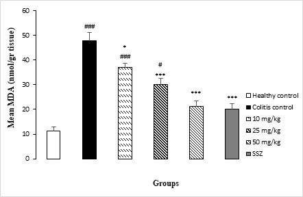
Figure 4: Effects of 3 different doses of D. kotschyi methanol extract and sulfasalazine (SSZ) on colonic MDA levels of colitis rats compared with healthy control and colitis control (CC) groups. *p < 0.05, ***p < 0.001 vs. colitis control; #P < 0.05, ###P < 0.001 vs. healthy control. Data are expressed as means ± SEM.

Figure 5: Effects of 3 different doses of D. kotschyi methanol extract and sulfasalazine (SSZ) on total thiol content in colonic tissue of colitis rats compared with healthy control and colitis control groups. *p < 0.05, **p <0.01 vs. colitis control; #p < 0.05 and ###p <0.001 vs. healthy control. Data are expressed as means ± SEM.
Table 1: Effects of various doses of D. kotschyi extract and sulfasalazine (SSZ) on colonic SOD and GPx activity in colitis rats compared with healthy and colitis control groups.
| Groups | Healthy control | Colitis control | D. kotschyi (10 mg/kg) |
D. kotschyi (25mg/kg) | D. kotschyi (50 mg/kg) |
SSZ | |||||
| SOD (u/mg) |
42.62±2.8 | 29.93±3# | 30.29±1.8# | 33.45±1.7 | 39.06±4.3 | 40.22±7.2 | |||||
| GPx (u/mg) |
359.4±34.1 | 136.3±21.8### | 210.7±99.4## | 225±10.6 | 306.7±23** | 276.7±25* | |||||
Note: *p < 0.05, **p < 0.01 vs. colitis control; #p < 0.05, ##p < 0.01 and ###p < 0.001 vs. healthy control. Data are expressed as means ± SEM
Acid secretion
Basal acid secretion in the healthy control rats averaged 7.2 ± 1.8 mEq/ml/15min period. In the rats in which colitis had been induced with acetic acid, basal acid secretion has a trend to increase but it was not statistically significant (data are not shown). D. kotschyi extract administration to the colitis rats did not induce any significant change in gastric acid secretion.
Discussion
The present study revealed that D. kotschyi induced improvements in various inflammatory parameters in an acetic acid-induced colitis model, which were shown through macroscopic, histological, and biochemical changes. Previous models of AA-induced colitis have been used for screening active drugs against inflammatory bowel disease and chemical phenomena related to the production of free radicals (22). In inflammatory conditions such as colitis, the natural balance between ROS production and the protective effect of free radical scavengers may be disturbed and resulted in tissue damage (23). Disturbed tissue protection makes the cells predispose to oxidative injury, which may contribute to the progression of the damage observed in experimental models of colitis (19). Therefore, increased oxidative stress and an impaired antioxidant defense likely play a role in UC pathogenesis.
Antioxidant enzymes such as SOD and GPx protect tissues against oxidative damage. Disorders in antioxidant defense system have been reported after colonic inflammation (24). The increase in tissue lipid peroxidation triggers a defective cycle that produces more and more metabolites that exhaust the antioxidant system of the cell and cause more inflammation (25). Therefore, it is logical to assume that treatment with D. kotschyi can improve the oxidative stress observed in experimental colitis because it reduced the MDA levels, which is an indicator of lipid peroxidation (24). In our study, MDA levels were reduced in treatment groups parallel to the macroscopic and histological changes. The decrease was more evident in D. kotschyi (50 mg/kg) and SSZ rats. These results suggest that D. kotschyi may have beneficial effects by reducing lipid peroxidation in damaged tissues of colon.
Sulfhydryl (SH), high-reactive components of protein molecules, are essential free radicals scavengers (26). It has revealed that SH groups are sensitive to oxidative injury and deplete after colitis induction (24). Our study showed a decrease in total sulfhydryl groups following the induction of colitis. Considering that the higher SH contents were observed in D. kotschyi treated rats than their respective control group, it is reasonable to suggest that D. kotschyi helped to replenish the total thiol pool.
One of the other important antioxidative defense mechanisms present in tissues is glutathione metabolism, which diminution that accelerates colon damages due to oxidative stress (25). In the current study, the data showed that GPx activity significantly decreased in control colitis group and treatment with D. kotschyi (50 mg/kg) restored that to the normal levels, suggesting an antioxidant effect of this plant. Therefore, an increase in colonic GPx activity may explain some of the beneficial effects of D. kotschyi in experimental colitis.
There are some reports of reduction of SOD activity in different studies as a result of oxidative stress (27). Superoxide anions can be catalyzed to H2O2 by scavenging the activity of SOD. In the present study, acetic acid administration reduced the SOD activity, which indicates the generation of reactive species (20, 27, 28), but no change in SOD activity was seen after the administration of D. kotschyi extract.
It has been shown that the D. kotschyi extract has a high amount of flavonoid and phenolic compounds, which have shown the highest antioxidant activity, and are probably responsible for the antioxidant activity of this plant (29, 30). The antioxidant activity can be mostly attributed to phenolic compounds such as luteolin, apigenin, xanthomicrol, rosmarinic acid and cirsimaritin in extracts of D. Kotschyi (31, 32). Phenolic compounds have redox potentials, which allow them to act as hydrogen donors and singlet oxygen quenchers.
The result of the present study showed that D. kotschyi extract exhibited antioxidant activity possibly through the modulation of antioxidant enzymes (GPx, and total thiol) as well as the suppression of lipid peroxidation. The findings of the current investigation support this notion that some medicinal plants are rich sources of natural antioxidants and flavonoids and could be used as therapeutic agents for some inflammatory diseases.
Since some studies have reported that chronic active gastritis is a common finding in patients with UC (33), we also performed a gastric acid secretion assay in our study. No change in basal gastric acid secretion was documented after colitis induction. Although a direct connection has been shown between the inflammation noted in the rectum and the stomach of IBD patients (34), the mechanisms underlying are poorly understood. Our results where consistent with the findings of some other studies (17), where no change in gastric acid secretion was shown after experimental colitis, which may suggest other contributing mechanisms in gastritis concomitant with the colitis.
Conclusion
In conclusion, our data indicate that the methanol extract of Dracocephalum kotschyi may diminish the colonic mucosal damage in experimental colitis by enhancing the activities of antioxidant enzymes and oxidative stress markers such as GPx and total thiol and by reducing the MDA (an oxidant indicator) level. Further investigations to clarify the mechanism of action of this plant extract is warranted.
Acknowledgements
A special thanks to Dr. Elham Amjadi for her technical assistance. Authors are also grateful to the Research Deputy of North Khorasan University of medical sciences for financial support to carry out this work.
Conflicts of Interest
There is no conflict of interest.
Received: 2020/10/12 | Accepted: 2021/01/25 | Published: 2021/10/17
| Rights and permissions | |
 |
This work is licensed under a Creative Commons Attribution-NonCommercial 4.0 International License. |
Copyright Policy
